
- Title
-
Embryonic and larval expression of zebrafish voltage-gated sodium channel alpha-subunit genes
- Authors
- Novak, A.E., Taylor, A.D., Pineda, R.H., Lasda, E.L., Wright, M.A., and Ribera, A.B.
- Source
- Full text @ Dev. Dyn.
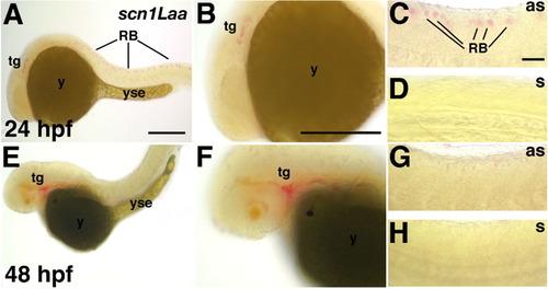
Scn1Laa is expressed in sensory neurons of the peripheral nervous system and Rohon-Beard (RB) cells. A-D: At 24 hours postfertilization (hpf), scn1Laa mRNA is detected in the trigeminal ganglion and RB cells. A: A low-magnification view reveals expression in an anterior domain caudal to the eye and in RB cells throughout the spinal cord. B: At higher magnification, the anterior expression domain is recognized as the developing trigeminal ganglion migrating rostrally toward the eye. C: The posterior expression is found in RB cells of the spinal cord. D: The sense probe (s) does not reveal a signal under the same conditions used for the antisense probe (as). E-H: At 48 hpf, in situ hybridization signals are stronger in the trigeminal ganglion but weaker posteriorly in the spinal cord. E,F: At 48 hpf, the trigeminal ganglia have migrated to their characteristic position just caudal to the eye and continue to express scn1Laa. G: At 48 hpf, dorsal RB cells continue to express scn1Laa transcripts. H: The sense probe reveals no hybridization signal. as, antisense probe; n, notochord; RB, Rohon-Beard cell; s, sense probe; tg, trigeminal ganglion; yse, yolk sac extension. In this and subsequent figures, whole-mount photos are oriented with anterior to the left and dorsal up. Scale bars = 250 μm in A (applies to A,E), 100 μm in B (applies to B,F), 100 μm in C (applies to C,D,G,H). |
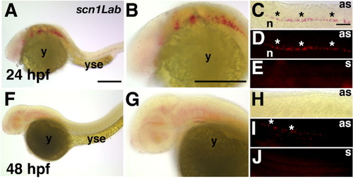
Scn1Lab expression is restricted to the central nervous system. A-E: At 24 hours postfertilization (hpf), scn1Lab transcripts are detected in ventral regions of the hindbrain and spinal cord. A: A low-magnification view reveals expression in the hindbrain and spinal cord. B: Scn1Lab expression is detected in ventral regions of the hindbrain. C: Similarly, in the spinal cord, scn1Lab expression is detected in ventral regions (asterisks). D: Epifluorescent illumination of the Fast Red in situ hybridization signal reveals expression ventrally within the spinal cord (asterisks). E: Using the same hybridization conditions as for antisense, the sense probe does not reveal a signal. F-J: At 48 hpf, scn1Lab expression is found diffusely within rostral regions of the central nervous system. F,G: Within the rostral central nervous system, scn1Lab expression is detected diffusely within ventral regions. H,I: Within the spinal cord, scn1Lab expression appears weaker at 48 versus 24 hpf because it is only detected by epifluorescent (I, asterisks) and not brightfield (H) illumination. J: The sense probe does not reveal a signal at 48 hpf, even using epifluorescent illumination. as, antisense probe; n, notochord; s, sense probe; y, yolk sac; yse, yolk sac extension. Scale bars = 250 μm in A (applies to A,F), 100 μm in B (applies to B,G), 100 μm in C (applies to C-E,H-J). |
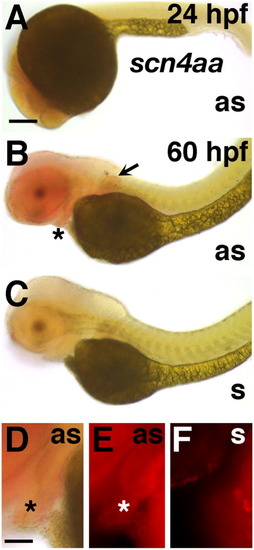
Scn4aa is expressed in mesodermal tissues. A: At 24 hours postfertilization (hpf), scn4aa mRNA is barely detected dorsal to the yolk sac. B: At 72 hpf, transcripts are detected in head muscle, pharyngeal muscle (asterisk), and the pectoral fin (arrow). C: The sense probe does not reveal a signal. D,E: At 72 hpf, pharyngeal muscle expresses scn4aa mRNA. F: The sense probe does not reveal expression. Scale bars = 250 μm in A (applies to A-C), 100 μm in D (applies to D-F). EXPRESSION / LABELING:
|
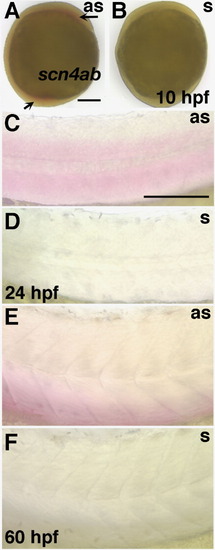
Scn4ab is expressed early in mesoderm and later in somatic tissue. A,B: At 10 hours postfertilization (hpf), scn4ab mRNA is detected in mesodermal tissue (arrows) using antisense but not sense probes. C,D: At 24 hpf, transcripts are detected in the trunk somites; in the dorsal somite, expression is most abundant in ventral region. E,F: At 60 hpf, expression is present in the somites and most abundantly in ventral half. Scale bars= 250 μm in A (applies to A,B), 100 μm in C (applies to C-F). |
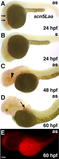
In situ hybridization reveals neural expression of scn5Laa. A: At 24 hours postfertilization (hpf), scn5Laa mRNA is detected in regions associated with the developing heart tube and pharynx (arrows). B: Under the same conditions, the sense probe does not reveal a signal. C: At 48 hpf, scn5Laa expression shifts to more anterior and dorsal regions of the embryo (e.g., head muscle, arrowhead). D,E: At 60 hpf, scn5Laa expression appears in rostral regions of the central nervous system as well as the developing pectoral fin (arrow). Scale bars = 250 μm in A (for A-E). as, antisense probe; s, sense probe. |

In situ hybridization reveals a temporally dynamic scn5Lab expression pattern. A-D: At 19 hours postfertilization (hpf), scn5Lab mRNA is detected in the developing somites of the trunk (asterisks in B,C). The in situ hybridization signal can be observed using either brightfield (B) or epifluorescent illumination (C). E: Under the same conditions, the sense probe does not reveal a signal. E-G: At 30 hpf, scn5Lab expression shifts from the peripheral somites to the central nervous system and is detected in ventral spinal cord (carats; E,F). G: The sense probe does not reveal a signal at 48 hpf, even using epifluorescent illumination. H-L: At 60 hpf, scn5Lab expression appears in the developing lateral line (I,L, arrows). In the trunk, expression is detected by epifluorescent (K) but not brightfield (J) illumination. L: The sense probe does not reveal a signal at 60 hpf, even using epifluorescent illumination. as, antisense probe; n, notochord; s, sense probe; yse, yolk sac extension. Scale bars = 250 and 100 μm in A (applies to A & H and K, respectively), 100 μm in B (applies to B-G,J-L). EXPRESSION / LABELING:
|
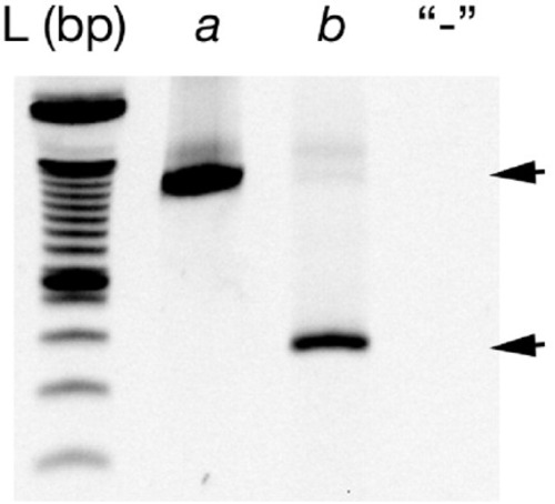
a,b: Reverse transcriptase-polymerase chain reaction (RT-PCR) reveals expression of both scn5Laa (a) and scn5Lab (b) in heart tissue of 72 hours postfertilization (hpf) embryos. The negative control (-) consisted of RT-PCR using an RT reaction from which the enzyme had been omitted. EXPRESSION / LABELING:
|
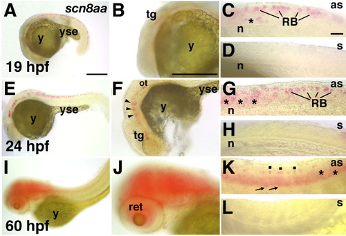
Scn8aa expression is restricted to the nervous system. A-E: At 19 hours postfertilization (hpf), scn8aa mRNA is detected in the developing trigeminal ganglion and spinal cord. A,B: In anterior portions of the embryo, scn8aa mRNA is detected in the developing trigeminal ganglion (tg). C: In the spinal cord, scn8aa transcripts are detected dorsally in Rohon-Beard (RB) cells as well as ventrally (asterisk). D: Under the same conditions, the sense probe does not reveal a signal. E-H: At 24 hpf, scn8aa expression expands within the hindbrain. E,F: scn8aa transcripts are detected in ventral hindbrain at 24 hpf (arrowheads) and the trigeminal ganglion. G: In the spinal cord, scn8aa expression occurs in RB cells and diffusely in ventral regions. H: The sense probe does not reveal a signal at 24 hpf. I-L: At 60 hpf, scn8aa expression appears diffusely in both rostral and caudal regions of the central nervous system. I,J: In rostral regions of the central nervous system, scn8aa expression is detected in the retina and diffusely in the brain. K: In the spinal cord, expression is detected dorsally in isolated cells (dots) and more diffusely in ventral regions (asterisks). In addition, expression is detected in the periphery, in dorsal root ganglia (arrows). L: The sense probe did not reveal a signal at 60 hpf. as, antisense probe; n, notochord; ot, otic vesicle; ret, retina; s, sense probe; y, yolk sac; yse, yolk sac extension. Scale bars = 250 μm in A (applies to A,E,I), 100 μm in B (applies to B,F,J), 100 μm in C (applies to C,D,G,H,K,L). EXPRESSION / LABELING:
|
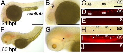
Scn8ab transcripts are detected in a subset of the scn8aa expression domain. A-E: At 24 hours postfertilization (hpf), scn8ab mRNA is detected in the developing trigeminal ganglion and some Rohon-Beard (RB) cells. A,B: In anterior portions of the embryo, scn8ab mRNA is detected in developing trigeminal ganglion. C,D: In the spinal cord, scn8ab transcripts are detected in some but not all RB cells. E: Under the same conditions, the sense probe does not reveal a signal. F-J: At 60 hpf, scn8ab expression expands to ventral spinal cord. F,G: In anterior regions, scn8ab transcripts are detected in the lateral line system (arrow). H,I: In the spinal cord, scn8ab expression persists in some RB cells and is now present diffusely in ventral regions (arrowheads). J: The sense probe does not reveal a signal at 60 hpf. as, antisense probe; n, notochord; s, sense probe; tg, trigeminal ganglion; y, yolk sac; yse, yolk sac extension. Scale bars-250 μm in A (applies to A,F), 250 μm in B (applies to B,H), 100 μm in C (applies to C-E,H-J). |

Unillustrated author statements EXPRESSION / LABELING:
|