
|
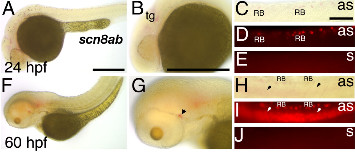
Fig. 10 Scn8ab transcripts are detected in a subset of the scn8aa expression domain. A-E: At 24 hours postfertilization (hpf), scn8ab mRNA is detected in the developing trigeminal ganglion and some Rohon-Beard (RB) cells. A,B: In anterior portions of the embryo, scn8ab mRNA is detected in developing trigeminal ganglion. C,D: In the spinal cord, scn8ab transcripts are detected in some but not all RB cells. E: Under the same conditions, the sense probe does not reveal a signal. F-J: At 60 hpf, scn8ab expression expands to ventral spinal cord. F,G: In anterior regions, scn8ab transcripts are detected in the lateral line system (arrow). H,I: In the spinal cord, scn8ab expression persists in some RB cells and is now present diffusely in ventral regions (arrowheads). J: The sense probe does not reveal a signal at 60 hpf. as, antisense probe; n, notochord; s, sense probe; tg, trigeminal ganglion; y, yolk sac; yse, yolk sac extension. Scale bars-250 μm in A (applies to A,F), 250 μm in B (applies to B,H), 100 μm in C (applies to C-E,H-J).