
- Title
-
Circadian time-keeping during early stages of development
- Authors
- Ziv, L., and Gothilf, Y.
- Source
- Full text @ Proc. Natl. Acad. Sci. USA
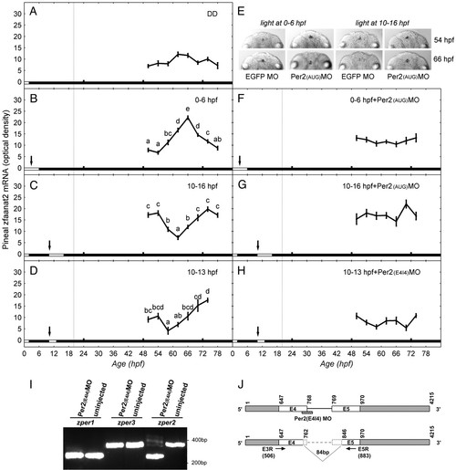
Effect of light and zper2 expression at early embryonic stage on development of pineal zfaanat2 mRNA rhythms. Zebrafish embryos, MO-injected and uninjected, were exposed to light for different periods during the first 16 h of development. During the third and fourth day of development, larvae were collected at 4-h intervals and pineal zfaanat2 mRNA levels were determined by ISH. (A) No rhythm was observed if embryos were placed in darkness at 2 hpf. (B–D) Rhythms of zfaanat2 mRNA appeared in embryos that were exposed to light 0–6 hpf (B), 10–16 hpf (C), or 10–13 hpf (D); note the phase difference induced by the light treatments. (F and G) Microinjection of Per2(AUG)MO at one-cell stage abolished the effect of the 0–6 hpf and 10–16 hpf light treatments (compare B to F and C to G). (H) Microinjection of Per2(E4I4)MO at one-cell stage abolished the effect of the 10–13 hpf light treatments (compare D to H). (E) Representative pineal zfaanat2 signals (dorsal view) at subjective midday (54 hpf) and subjective midnight (66 hpf) of embryos that were exposed to light at 0–6 hpf (Left) or 10–16 hpf (Right) and were injected with Per2(AUG)MO or EGFP MO. Each value represents the average optical density of pineal signal ± SEM in 20–60 embryos. Different letters represent statistically different values (P < 0.05, ANOVA, Tukey’s test). The horizontal bar at the bottom of each panel represents the lighting conditions: white boxes represent the illumination period, and black boxes represent dark. Arrows indicate the beginning of effective illumination. Vertical dashed lines represent the time of pineal gland formation (i.e., earliest detection of photoreceptor cells). (I and J) Per2(E4I4)MO injection altered zper2 mRNA splicing but had no effect on the splicing of zper1 and zper3, as revealed by PCR analysis (I). Sequence analysis of the PCR products indicate that Per2(E4I4)MO caused the skipping of 84 nucleotides in exons 4 and 5 (7 bp in exon 4 and 77 bp in exon 5), which encode a 28-aa fragment in zPER2 PAS A domain (J). EXPRESSION / LABELING:
PHENOTYPE:
|
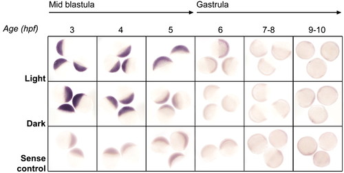
Effect of light on zper2 expression at the blastula and gastrula stages. Embryos were kept under continuous light or dark and sampled throughout the first 10 h of development; zper2 mRNA was detected by whole-mount ISH. Shown are representative whole-mount ISH results of 3–10-h zebrafish embryos, side view; the embryonic stage and age are indicated at the top. Before transcription activation at the MBT stage (3–4 hpf), maternal zper2 mRNA was present at high levels in all embryos regardless of the light treatment. Under continuous dark, zper2 transcripts rapidly decreased, and by 5 hpf, the signal was similar to that obtained with the sense RNA control probe. Under continuous light, the decay in zper2 mRNA was substantially slower; the signal matched that obtained with sense RNA probe at 7–8 hpf. EXPRESSION / LABELING:
|
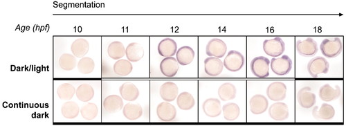
Effect of light on zper2 expression during segmentation period. Embryos were kept under constant darkness (Lower) or exposed to light 10–16 hpf (Upper), sampled at 1-h intervals during 10–18 hpf, and zper2 mRNA was detected by whole-mount ISH. Shown are representative whole-mount ISH results of 10- to 18-hpf embryos, lateral view. Zebrafish per2 mRNA was undetectable in embryos kept under dark conditions (Lower), whereas exposure to light resulted in de novo transcription of zper2 (Upper). Zebrafish per2 mRNA levels remained high throughout the illumination period and declined only after lights-off. The horizontal bar at the bottom of each panel represents the lighting conditions before and during sampling; white boxes represent light and black boxes represent dark. EXPRESSION / LABELING:
|

Spatio-temporal expression pattern of zper2 during neurogenesis. Whole-mount ISH analysis of zper2 mRNA in 12- to 22-h embryos after light treatment (dorsal view, anterior to the top) is shown. (A) Enhanced zper2 mRNA expression is detected at the lateral edge of the anterior neural plate (black arrows) and the median region of the neural plate (white arrow) beginning at the 6-somite-stage (12 hpf). (B and C) Expression of zper2 at the pineal gland primordium (black arrowhead) begins at the 18-somite stage (18 hpf, B) and is further enhanced at the 24-somite stage (C). At this stage, enhanced zper2 expression also occurs in the eye area. s, somites. EXPRESSION / LABELING:
|