
- Title
-
Roles for fgf8 signaling in left-right patterning of the visceral organs and craniofacial skeleton
- Authors
- Albertson, R.C., and Yelick, P.C.
- Source
- Full text @ Dev. Biol.
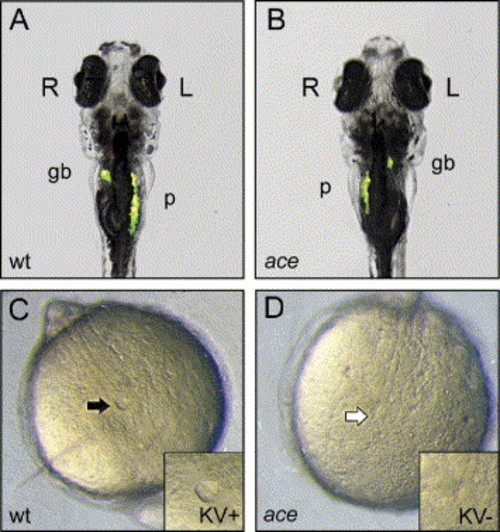
fgf8 is required for LR asymmetric development of the viscera and morphogenesis of Kupffer′s vesicle (KV). (A–B) By 7 dpf the gall bladder (gb) and pancreas (p) autofluorescence under UV illumination, providing a simple screen for LR situs defects. (A) Wt larvae typically exhibit normal LR situs positioning (situs solus), with the gall bladder to the right of the midline and the pancreas to the left. (B) Approximately 30% of ace/fgf8 mutants exhibit randomized LR situs positioning, including situs inversus. (C) KV is apparent in wt embryos by the five somate stage (black arrow). (D) KV does not develop in approximately 30% of ace/fgf8 mutants (white arrow where KV should be). |
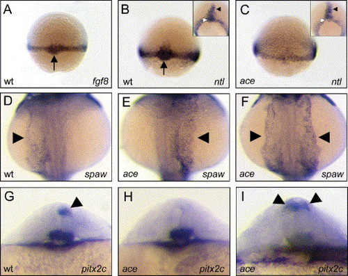
(A, B) At shield stage, both fgf8 and ntl mRNAs are expressed in wt dorsal forerunner cells DFCs (black arrows). (C) ntl expression is reduced in the margin and DFCs of age-matched ace/fgf8 mutants. (B, C insets) By ∼70% epiboly ntl expression in the midline of ace/fgf8 mutants is indistinguishable from that in age-matched wt embryos (black arrowheads). At this stage, mutant embryos can still be distinguished from wts by reduced ntl expression in DFCs (white arrowheads). (D–F) fgf8 deficiency results in randomization of nodal signaling in the lateral plate mesoderm (LPM) (arrowheads). (D) The nodal-related gene spaw is expressed in the left LPM of wt embryos at 22 somites. Similarly staged ace/fgf8 mutants exhibit reversed (E) and bilaterally (F) expressed spaw in the LPM. (G–I) Normal LR asymmetric development of the diencephalon is disrupted in ace/fgf8 mutants (arrowheads). (G) At 26 hpf, pitx2c is expressed on the left side of the dorsal diencephalon. Expression of this marker is either absent (H) or bilateral (I) in ace/fgf8 mutants. EXPRESSION / LABELING:
|
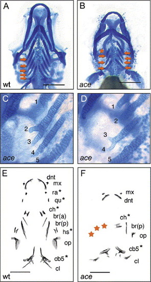
Asymmetric craniofacial development in ace/fgf8 mutants. All panels display a ventral view of the pharyngeal skeleton, with the right side of the pharynx present on the left, and the left side present on the right. (A) At 5 dpf, wt larvae exhibit five sets of paired bilateral cb cartilages (orange arrows). (B) Mutant larvae exhibit reduced and asymmetric numbers of cb cartilages (asterisk indicates position of asymmetrically missing cartilage). Pharyngeal cartilage asymmetry in ace/fgf8 mutants has a directional bias with elements typically missing from the right side of the pharynx (Χ2 = 14.5, P < 0.001). (C, D) ace/fgf8 mutants exhibit lateral branching of various cb cartilages. (E, F) ace/fgf8 mutants exhibit asymmetric pharyngeal bone development. (E) By 7 dpf, wt larvae possess a bilateral series of 11 mineralized bones. Pharyngeal bones are labeled to the right side of the figure (see abbreviations below). Starred bones represent cartilage replacement bones; all others are dermal bones. (F) Mutant larvae exhibit reduced and asymmetric bone development with a bias toward the right side of the pharynx (Χ2 = 3, P = 0.08) (red asterisks indicate position of asymmetrically missing bones). Abbreviations are as follows: br(a), anterior branchiostegal ray; br(p), posterior branchiostegal ray; cb5, fifth ceratobranchial cartilage; ch, ceratohyal; cl, cleithrum; dnt, dentary; hs, hyosymplectic; mx, maxilla; op, opercle; qu, quadrate; ra, retroarticular. |
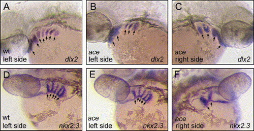
ace/fgf8 mutants exhibit asymmetric pharyngeal arch development. (A–C) Cranial neural crest (CNC) segments at 36 hpf in wt and mutant larvae (black arrows). (A) Wt dlx2 expression in 7 bilateral CNC segments. (B, C) Age-matched ace/fgf8 mutants exhibit fewer and often asymmetrical numbers of CNC segments. (D–F) Pharyngeal endoderm pouches in wt and mutant larvae (black arrows). (D) At 32 hpf, wt larvae possess a bilateral series of pharyngeal endodermal pouches, revealed via nkx2.3 expression. (E, F) In some ace/fgf8 mutants, the pharyngeal endoderm develops asymmetrically, with fewer endodermal pouches apparent on the right side of the pharynx. |
Reprinted from Developmental Biology, 283(2), Albertson, R.C., and Yelick, P.C., Roles for fgf8 signaling in left-right patterning of the visceral organs and craniofacial skeleton, 310-321, Copyright (2005) with permission from Elsevier. Full text @ Dev. Biol.