
- Title
-
Distinct tissue-specificity of three zebrafish ext1 genes encoding proteoglycan modifying enzymes and their relationship to somitic Sonic Hedgehog signaling
- Authors
- Siekmann, A.F., and Brand, M.
- Source
- Full text @ Dev. Dyn.
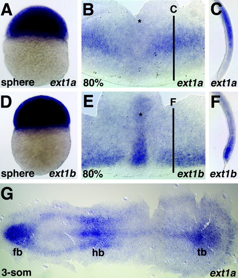
Figure 2. Expression of ext1a (A-C,G) and ext1b (D-F), anterior to the top (A-F), or to the left (G). A: Sphere stage. B: Eighty percent of epiboly. The embryo was opened at the ventral side and flat-mounted, dorsal side up. Ext1a transcripts can be detected in two wings in the prospective hindbrain region lateral to the embryonic midline (marked by asterisk). C: Cross-section of the embryo shown in B. D: Sphere stage. E: Eighty percent epiboly stage. Orientation of the embryo as in B. Ext1b RNA can be detected in the embryonic midline (marked by asterisk) and in the germ ring. F: Cross-section of the embryo in E. G: At the three-somite (3-som) stage, ext1a is expressed in the forebrain (fb), the hindbrain (hb), and in the tailbud (tb). EXPRESSION / LABELING:
|
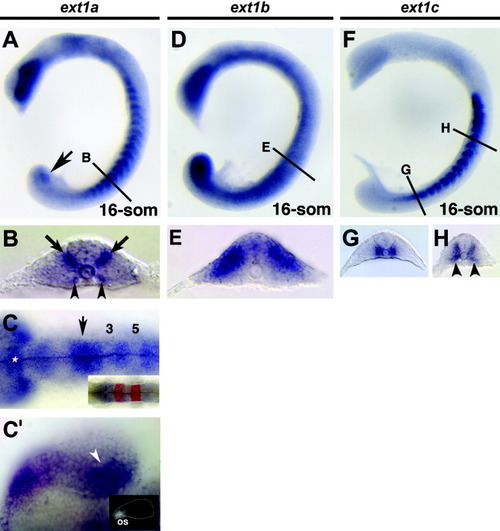
Figure 3. Expression of ext1a (A-C,C), ext1b (D,E), and ext1c (F-H) at the 16-somite stage. Anterior of the embryos is to the left. A: Expression of ext1a can be detected in the eye, the dorsal neural tube, Kupffer′s vesicle (black arrow), and the somites. B: Cross-section at the level of the 12th somite. Expression of ext1a can be detected in the anterior hindbrain (arrow) and the dorsal diencephalon (asterisk). C: Dorsal view on the brain of the embryo in A. Krox20 demarcates rhombomeres 3 and 5 (inset). C: Expression of ext1a in the eye, which do not express ext1a. In addition, ext1a transcripts can be detected in two ventromedial (arrowheads) and two dorsomedial (arrows) domains. D: RNA encoding ext1b is expressed more ubiquitously with domains of higher expression in the tailbud region, the eye, and dorsal somites. E: Section at the level of the 9th somite, revealing expression of ext1b in the dorsal aspect of the somite. F: Alternating expression domains of ext1c can be detected in the somites. G: Cross-section of the embryo in F at the level of the 16th somite. H: Cross-section at the level of the 6th somite, revealing expression of ext1c in ventromedial regions of the somites (arrowheads) and adjacent to the neural tube. EXPRESSION / LABELING:
|

Figure 4. Expression of ext1a (A-F), ext1b (G-J), and ext1c (K-O) at 24 hr postfertilization (hpf) (A-G,I-K,M) and 48 hpf (B,H,L,N,O). A: Neural expression of ext1a can be detected in the cerebellum (arrow), the dorsal diencephalon (asterisk) and the optic stalk (arrowhead). B: Expression in the otic vesicle (black arrow). C: Cross-section of the embryo in A at the level of the anterior spinal cord. D: Dorsal view of the embryo in A at the level of the cerebellum (black arrow) and hindbrain. Ov, otic vesicle. E: Dorsal diencephalon (outlined with white dots; A, anterior, P, posterior). F: Cross-section through the eye. Black arrowhead marks optic stalk. G: Expression of ext1b in the tailbud region (arrowhead) and in anterior neural tissue (arrow). H: The fins (black arrow) express ext1b. I: Close up of cells belonging to the anterior pronephric duct (black arrow). J: Close up of the fin bud mesenchyme. K: Transcripts of ext1c can be detected in the telencephalon (black arrow) and in the rhombomeres (white arrowhead). L: Expression is confined to the brain. M: Telencephalon (white asterisk) and the olfactory bulbs (marked by arrow and outlined by dotted white line) of the embryo in A show ext1c expression. N: Expression of ext1c in the retina. O: Cross-section of the embryo in L at the level of the otic vesicles (white arrowhead). The white matter is marked by black arrow. |

Figure 5. Dependence of ext1a and ext1c expression on Sonic hedgehog (Shh) signaling. A-F: Dorsal views of 14-somite stage embryos with anterior to the left. Expression of ext1a (A-C) and ext1c (D-F), respectively. A,B,D,E: In smu mutant embryos, expression of ext1a (A) and ext1c (D) is greatly reduced compared with wild-type (wt) embryos (B,E). C,F: Overexpression of shh results in an expansion of the somitic expression domains of ext1a (C) and ext1c (F, compare white brackets in B,C and E,F). Black lines indicate somite borders in (A-C). EXPRESSION / LABELING:
|