
- Title
-
Identification of a nonchordate-type classic cadherin in vertebrates: Chicken Hz-cadherin is expressed in horizontal cells of the neural retina and contains a nonchordate-specific domain complex
- Authors
- Tanabe, K., Takeichi, M., and Nakagawa, S.
- Source
- Full text @ Dev. Dyn.
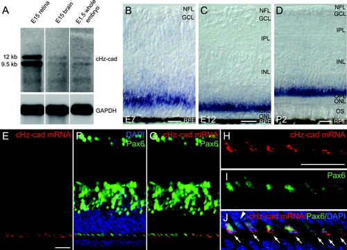
Chicken Hz-cadherin (cHz-cadherin) is expressed in horizontal cells of the chicken neural retina. A: Northern-blot analysis of the expression of cHz-cadherin mRNA. B-D: In situ hybridization of embryonic day 7 (E7; B), E12 (C), and posthatching chick retina (D) probed with cHz-cadherin. Note that the blue color reaction products of the alkaline phosphatase tend to diffuse and strong signals were observed in the outer plexiform layer as well as the cell bodies located in the outer most region of the inner nuclear layer. E-J: Simultaneous detection of cHz-cadherin mRNA (green in F,G,I,J) and horizontal cell marker Pax6 (red in E,G,H,J) on the same sections of the E12 retina using confocal microscopy. Counter nuclear staining of DAPI (4′,6-diamidine-2-phenylidole-dihydrochloride) is shown in blue in F and J. H-J show higher magnification of the outer region of the retina shown in E-G. Arrows point to the cells coexpressing cHz-cadherin mRNA and Pax6, and the arrowhead shows the cells solely expressing Pax6. NFL, neurofilament layer; GCL, ganglion cell layer; IPL, inner plexiform layer; INL, inner nuclear layer; OPL, outer plexiform layer; ONL, outer nuclear layer; OS, outer segment; RPE, retinal pigment epithelium. Scale bars = 20 μm in B-D, E (applies to E-G), H (applies to H-J). |
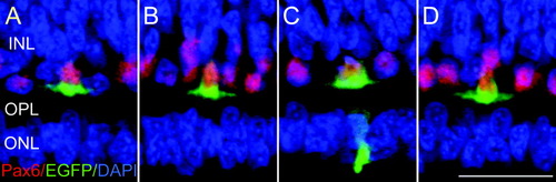
Pax6-expressing cells in the outer portion of the inner nuclear layer are horizontal cells. A-D: Confocal images of the outer portion of an embryonic day 12 neural retina stained with Pax6 (red), electroporated green fluorescence protein (EGFP; green), and DAPI (blue). Four horizontal cells expressing GFP were randomly selected. INL, inner nuclear layer; OPL, outer plexiform layer; ONL, outer nuclear layer. Scale bar = 20 μm in D (applies to A-D). |
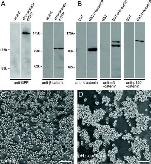
Chicken Hz-cadherin (cHz-cadherin) functions as a cell adhesion molecule. A: Expression of cHz-cadherin and stabilization of β-catenin in the A431D transfectants. A431D cells were stably transfected with cHz-cadherin tagged with GFP, and the transgene expressions were examined by Western blotting using anti-GFP and anti-β-catenin monoclonal antibody. Note the control A431D cells express little β-catenin, whereas this protein was stabilized upon the introduction of cHz-cadherin. B: In vitro binding of catenins with the cytoplasmic domain of cHz-cadherin. Whole brain lysates were incubated with control (GST alone) or cHz-cadherin-GST fusion protein, and bound proteins were separated by sodium dodecyl sulfate-polyacrylamide gel electrophoresis and detected with the antibodies shown below. C,D: Aggregation of control A431D cells (C) and cHz transfectants (D). cHz-cadherin-expressing cells form cellular aggregates, whereas most of the parental A431D cells remain as single cells. Scale bars = 50 μm in C,D. |