
Fig. 2
- ID
- ZDB-FIG-061122-30
- Publication
- Tanabe et al., 2004 - Identification of a nonchordate-type classic cadherin in vertebrates: Chicken Hz-cadherin is expressed in horizontal cells of the neural retina and contains a nonchordate-specific domain complex
- Other Figures
- All Figure Page
- Back to All Figure Page
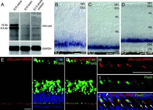
Chicken Hz-cadherin (cHz-cadherin) is expressed in horizontal cells of the chicken neural retina. A: Northern-blot analysis of the expression of cHz-cadherin mRNA. B-D: In situ hybridization of embryonic day 7 (E7; B), E12 (C), and posthatching chick retina (D) probed with cHz-cadherin. Note that the blue color reaction products of the alkaline phosphatase tend to diffuse and strong signals were observed in the outer plexiform layer as well as the cell bodies located in the outer most region of the inner nuclear layer. E-J: Simultaneous detection of cHz-cadherin mRNA (green in F,G,I,J) and horizontal cell marker Pax6 (red in E,G,H,J) on the same sections of the E12 retina using confocal microscopy. Counter nuclear staining of DAPI (4′,6-diamidine-2-phenylidole-dihydrochloride) is shown in blue in F and J. H-J show higher magnification of the outer region of the retina shown in E-G. Arrows point to the cells coexpressing cHz-cadherin mRNA and Pax6, and the arrowhead shows the cells solely expressing Pax6. NFL, neurofilament layer; GCL, ganglion cell layer; IPL, inner plexiform layer; INL, inner nuclear layer; OPL, outer plexiform layer; ONL, outer nuclear layer; OS, outer segment; RPE, retinal pigment epithelium. Scale bars = 20 μm in B-D, E (applies to E-G), H (applies to H-J). |