
Fig. 4
- ID
- ZDB-FIG-061122-32
- Publication
- Tanabe et al., 2004 - Identification of a nonchordate-type classic cadherin in vertebrates: Chicken Hz-cadherin is expressed in horizontal cells of the neural retina and contains a nonchordate-specific domain complex
- Other Figures
- All Figure Page
- Back to All Figure Page
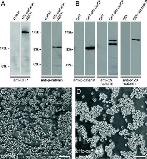
Chicken Hz-cadherin (cHz-cadherin) functions as a cell adhesion molecule. A: Expression of cHz-cadherin and stabilization of β-catenin in the A431D transfectants. A431D cells were stably transfected with cHz-cadherin tagged with GFP, and the transgene expressions were examined by Western blotting using anti-GFP and anti-β-catenin monoclonal antibody. Note the control A431D cells express little β-catenin, whereas this protein was stabilized upon the introduction of cHz-cadherin. B: In vitro binding of catenins with the cytoplasmic domain of cHz-cadherin. Whole brain lysates were incubated with control (GST alone) or cHz-cadherin-GST fusion protein, and bound proteins were separated by sodium dodecyl sulfate-polyacrylamide gel electrophoresis and detected with the antibodies shown below. C,D: Aggregation of control A431D cells (C) and cHz transfectants (D). cHz-cadherin-expressing cells form cellular aggregates, whereas most of the parental A431D cells remain as single cells. Scale bars = 50 μm in C,D. |