
Image
|
Figure Caption
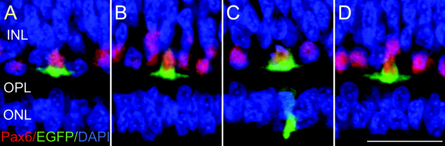
Fig. 3 Pax6-expressing cells in the outer portion of the inner nuclear layer are horizontal cells. A-D: Confocal images of the outer portion of an embryonic day 12 neural retina stained with Pax6 (red), electroporated green fluorescence protein (EGFP; green), and DAPI (blue). Four horizontal cells expressing GFP were randomly selected. INL, inner nuclear layer; OPL, outer plexiform layer; ONL, outer nuclear layer. Scale bar = 20 μm in D (applies to A-D).
Acknowledgments
This image is the copyrighted work of the attributed author or publisher, and
ZFIN has permission only to display this image to its users.
Additional permissions should be obtained from the applicable author or publisher of the image.
Full text @ Dev. Dyn.