
- Title
-
Cardiac neural crest contributes to cardiomyogenesis in zebrafish
- Authors
- Sato, M. and Yost, H.J.
- Source
- Full text @ Dev. Biol.
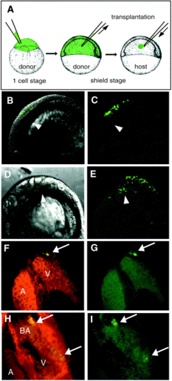
Transplanted neural crest cells transform into muscle cells in the myocardium. (A) Diagram of cell transplantation procedure. Alexa Fluor 488 was injected into donor embryos at the 1-cell stage. At the shield stage, donor cells were transplanted at the position of neural crest to the homotypic position of an age-matched host. This position does not overlap with the ventral and equatorial mesoderm that will give rise to the heart. (B–E) Lateral view, cranial toward left. DIC and fluorescent images are superimposed in left panels, and fluorescent image in right panels. (B, C) At the 8-somite stage, embryos were selected that had transplanted cells (green label) in the neural crest, not in the lateral plate mesoderm or heart-forming precardiac regions (arrowhead). (D, E) At the 26-somite stage, labeled neural crest cells have migrated toward the heart-forming regions (arrow head). (F–I) Confocal cross sections of the heart, dorsal view, cranial to the top. Green (lineage-label) and red channel (MF20 immunofluorescence) overlap in left panels. Lineage-labeled cells were shown in right panels. (F, G) At 3 dpf, in the same embryo as shown above, a lineage-labeled transplanted cell (green label and arrow, G) was detected in the ventricular (V) myocardium, and was MF20-positive (yellow label in overlapped images, F). A indicates the atrium. (H, I) In another embryo, transplanted neural crest cells (arrows) were detected in the bulbus arteriosus (BA) and ventricle (V) and were also MF20-positive (yellow labels in H). Myocardial nuclei are not stained by MF20. |
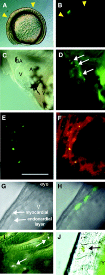
Two lineage-label approaches mark cardiac neural crest cells that form muscle cells in the myocardium. (A–F, I) Laser uncaging of DMNB-caged fluorescein dextran. Normarski optics (A) and epifluorescence (B) lateral view of an 8-somite-stage embryo in which 15 cells in the neural crest were labeled by laser uncaging (green in B). Yellow arrowheads show cardiac neural crest region in chick. (C, D) Lateral views of the heart in 72-hpf embryo with cranial to the left and dorsal up. Normarski optics (C) and epifluorescence view (D). Arrows indicate labeled cells in the bulbus arteriosus (BA), the ventricle (V), and the atrioventricular junction between the ventricle and atrium (A). Autofluorescence was seen in yolk cells (Y) but not in cells of the embryo. (E, F) Confocal images of the ventricle of a 72-hpf embryo. Green channel (lineage label) shown in left panel, superimposed green and red (MF20) channels shown in right panel. Labeled neural crest cells (green in E) were detected as MF20-positive cells (double STAINING = yellow in F) embedded in a field of MF20-positive (red) ventricle cardiomyocytes. By 72 hpf, the lineage-labeling begins to appear punctate within the cytoplasm of lineage-labeled cells. Scale bar, 20 μm. (G, H, J) Laser activation of hsp70-gfp transgene in neural crest lineages resulted in GFP-labeled myocytes at 36 hpf. Lateral views of the heart with cranial to the top and ventral left. Both myocardial layer and endocardial layer in the ventricle were detected in Normarski optics (G). Three fluorescent-labeled cells were detected in the myocardial layer (H). (I, J) Cells labeled by laser uncaging or GFP-activation were also detected in (I) the pharyngeal arches and head cartilage and (J) a pigment cell, indicating that these techniques successfully labeled neural crest cells. Arrows indicate labeled cells. |
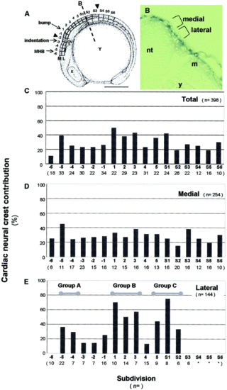
Cardiac neural crest originates from a broad rostrocaudal region in zebrafish. (A) Topological map used for fate mapping. A schematic drawing of an 8-somite embryo shown in a lateral view with cranial to the left and dorsal up (modified from Kimmel et al., 1995). Neural crest were divided rostrocaudally into 17 divisions designated -6 through S6. Arrowheads show cardiac neural crest area in chick. Each division was further subdivided into medial (M) and lateral (L) layers along the orthogonal axis. Line B indicates region of cross section in (B). Y, yolk; E, eye; OV, otic vesicle; MHB, midbrain/hindbrain boundary; M, medial; L, lateral subdivision; scale bar, 250 μm. (B) Cross section of the 8-somite embryo, indicating medial and lateral neural crest populations with respect to the neural tube (nt), lateral mesoderm (m), and yolk (y). (C–E) Each subdivision was labeled in separate embryos, and percent of embryos in which labeled cardiac neural crest cells were detected in the heart was scored. Scores within each rostrocaudal division were combined (C) or presented separately for the medial (D) and lateral (E) subdivisions. Bold numbers and letters below each graph correspond with the rostrocaudal topological map position. Lower numbers, in parenthesis, indicate the number of appropriately labeled embryos. Lateral neural crest from 3 regions (Groups A, B, and C) displayed large contributions to the heart. In lateral subdivision positions -6 and S3, labeled neural crest did not contribute to the heart. In divisions S4-S6, it was difficult to define lateral neural crest subdivisions because of the developing somites (*). |

Distinct gene expression patterns in three cardiac neural crest groups. (A–G) Lateral views of embryos at the eight-somite stage; cranial to the left and dorsal up. (A) Arrowheads in the diagram show the fate-map boundaries of the three cardiac neural crest groups along rostrocaudal axis: Group A (red), Group B (green), Group C (blue). (B–G) Whole-mount in situ hybridization of neural crest markers indicated a bottom of each panel. (H) Summary of neural crest gene expression patterns. The cardiac neural crest fate-map groups in which these genes are expressed are indicated in gray in the chart. wnt1 and wnt3a are not expressed in group A, and lef1 is weakly expressed. wnt3a is only expressed in group B. |
Reprinted from Developmental Biology, 257(1), Sato, M. and Yost, H.J., Cardiac neural crest contributes to cardiomyogenesis in zebrafish, 127-139, Copyright (2003) with permission from Elsevier. Full text @ Dev. Biol.