
Fig. 2
- ID
- ZDB-FIG-080423-33
- Publication
- Sato et al., 2003 - Cardiac neural crest contributes to cardiomyogenesis in zebrafish
- Other Figures
- All Figure Page
- Back to All Figure Page
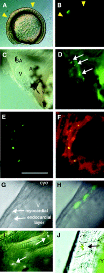
Two lineage-label approaches mark cardiac neural crest cells that form muscle cells in the myocardium. (A–F, I) Laser uncaging of DMNB-caged fluorescein dextran. Normarski optics (A) and epifluorescence (B) lateral view of an 8-somite-stage embryo in which 15 cells in the neural crest were labeled by laser uncaging (green in B). Yellow arrowheads show cardiac neural crest region in chick. (C, D) Lateral views of the heart in 72-hpf embryo with cranial to the left and dorsal up. Normarski optics (C) and epifluorescence view (D). Arrows indicate labeled cells in the bulbus arteriosus (BA), the ventricle (V), and the atrioventricular junction between the ventricle and atrium (A). Autofluorescence was seen in yolk cells (Y) but not in cells of the embryo. (E, F) Confocal images of the ventricle of a 72-hpf embryo. Green channel (lineage label) shown in left panel, superimposed green and red (MF20) channels shown in right panel. Labeled neural crest cells (green in E) were detected as MF20-positive cells (double STAINING = yellow in F) embedded in a field of MF20-positive (red) ventricle cardiomyocytes. By 72 hpf, the lineage-labeling begins to appear punctate within the cytoplasm of lineage-labeled cells. Scale bar, 20 μm. (G, H, J) Laser activation of hsp70-gfp transgene in neural crest lineages resulted in GFP-labeled myocytes at 36 hpf. Lateral views of the heart with cranial to the top and ventral left. Both myocardial layer and endocardial layer in the ventricle were detected in Normarski optics (G). Three fluorescent-labeled cells were detected in the myocardial layer (H). (I, J) Cells labeled by laser uncaging or GFP-activation were also detected in (I) the pharyngeal arches and head cartilage and (J) a pigment cell, indicating that these techniques successfully labeled neural crest cells. Arrows indicate labeled cells. |
Reprinted from Developmental Biology, 257(1), Sato, M. and Yost, H.J., Cardiac neural crest contributes to cardiomyogenesis in zebrafish, 127-139, Copyright (2003) with permission from Elsevier. Full text @ Dev. Biol.