Fig. 3
- ID
- ZDB-FIG-080423-34
- Publication
- Sato et al., 2003 - Cardiac neural crest contributes to cardiomyogenesis in zebrafish
- Other Figures
- All Figure Page
- Back to All Figure Page
|
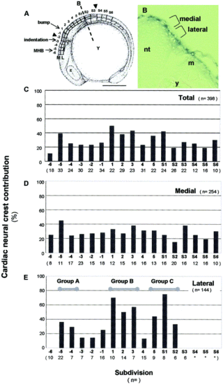
Cardiac neural crest originates from a broad rostrocaudal region in zebrafish. (A) Topological map used for fate mapping. A schematic drawing of an 8-somite embryo shown in a lateral view with cranial to the left and dorsal up (modified from Kimmel et al., 1995). Neural crest were divided rostrocaudally into 17 divisions designated -6 through S6. Arrowheads show cardiac neural crest area in chick. Each division was further subdivided into medial (M) and lateral (L) layers along the orthogonal axis. Line B indicates region of cross section in (B). Y, yolk; E, eye; OV, otic vesicle; MHB, midbrain/hindbrain boundary; M, medial; L, lateral subdivision; scale bar, 250 μm. (B) Cross section of the 8-somite embryo, indicating medial and lateral neural crest populations with respect to the neural tube (nt), lateral mesoderm (m), and yolk (y). (C–E) Each subdivision was labeled in separate embryos, and percent of embryos in which labeled cardiac neural crest cells were detected in the heart was scored. Scores within each rostrocaudal division were combined (C) or presented separately for the medial (D) and lateral (E) subdivisions. Bold numbers and letters below each graph correspond with the rostrocaudal topological map position. Lower numbers, in parenthesis, indicate the number of appropriately labeled embryos. Lateral neural crest from 3 regions (Groups A, B, and C) displayed large contributions to the heart. In lateral subdivision positions -6 and S3, labeled neural crest did not contribute to the heart. In divisions S4-S6, it was difficult to define lateral neural crest subdivisions because of the developing somites (*). |
Reprinted from Developmental Biology, 257(1), Sato, M. and Yost, H.J., Cardiac neural crest contributes to cardiomyogenesis in zebrafish, 127-139, Copyright (2003) with permission from Elsevier. Full text @ Dev. Biol.

