
- Title
-
B cells develop in the zebrafish pancreas
- Authors
- Danilova, N. and Steiner, L.A.
- Source
- Full text @ Proc. Natl. Acad. Sci. USA
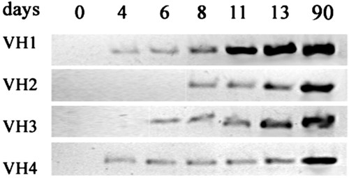
Time course of Igμ gene rearrangement during development. Total genomic DNA from about 30 larvae was subjected to PCR with primers specific for each of the VH families indicated. Amplification occurred only after VDJ rearrangement of genes in that family. |
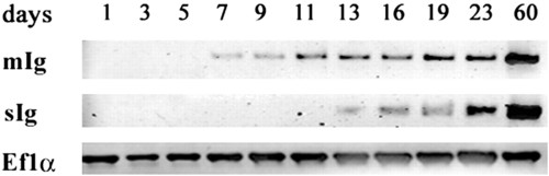
Expression of membrane (mIg) and secreted (sIg) forms of Igμ during development, estimated by RT-PCR. Total RNA was prepared from 50-100 fish at each time point. PCR was performed with an upstream primer, Cμ3S, corresponding to a segment of the CH3 domain, which is found in both the secreted and membrane forms of Igμ; the downstream primers, Ctail and tm2, are specific for the secreted and membrane carboxyl-terminal tail pieces, respectively. The expression of elongation factor 1α served as a standard. |
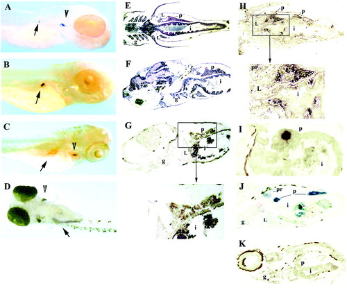
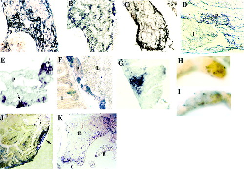
Expression of Igμ and rag1 during development. Whole-mount in situ hybridization of 4-day fish with rag1 (A) and insulin (B) probes. In addition to strong staining with the rag1 probe in the thymus (A, arrowhead), weak staining can be detected in the right dorsal aspect of the abdominal cavity (A, arrow), where the developing pancreas is located; the location of staining with the insulin probe (B, arrow) appears to coincide with that by the rag1 probe. (C) Whole-mount in situ hybridization of 5-day fish with the rag1 probe. The arrowhead points to the thymus and the arrow to the pancreas; the central unstained area within the pancreas corresponds to an islet of Langerhans. (D) Whole-mount in situ hybridization of 8-day fish with rag1 probe, ventral view. Staining is seen in thymus (arrowhead) and pancreas (arrow); the pancreas is now elongated. Horizontal (E) and sagittal (F) paraffin sections through a 10-day-old fish stained with hematoxylin/eosin. The intestine at this age is a straight tube; the pancreas extends along the intestine. In situ hybridization of sagittal (G) or horizontal (H and I) cryosections of 10-day fish with Igμ (G), rag1 (H), or insulin (I) probes. Staining with rag1 and Igμ probes is seen in the pancreas surrounding an islet of Langerhans; with the rag1 probe, a few intraepithelial cells within the intestine also are stained. Intense staining of an islet is seen with the insulin probe. (J) In situ hybridization of a sagittal cryosection of 19-day fish with Igμ probe; most staining is within the pancreas, but some is also seen in the pronephros. (K) In situ hybridization of sagittal cryosection with Igμ sense probe; the brown color is pigment. L, liver; g, gills; i, intestine; p, pancreas; pr, pronephros. The fish in A is albino; those in B-D are wild-type treated with phenylthiourea and the rest are untreated wild-type. The dark material within the intestine in G-J is remnants of food. |
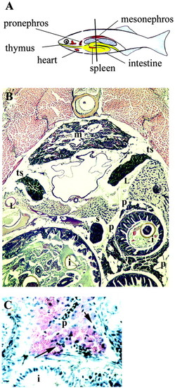
Organs of adult zebrafish. (A) Diagram illustrating the position of some of the organs; the liver, pancreas, and gonads are not shown. The liver wraps around the anterior part of the intestine. Pancreatic tissue, intermixed with lymphatic tissue, is dispersed along the intestine. The bilateral gonads extend from the central to the posterior part of the abdominal cavity. The vertical line indicates the position of the section shown in B. (B) Transverse paraffin section, hematoxylin/eosin stain. Three crosssections of intestine (i) are seen; segments of pancreatic tissue (p) are located along the intestine. The mesonephros (m) contains renal tubules and some hematopoietic tissue. The spleen (s) is a compact organ containing, almost exclusively, erythrocytes. Bilateral testes (t) are located adjacent to the swim bladder. Cross sections of liver (L) are also seen. (C) Higher magnification of section similar to that in B, showing wall of intestine and adjacent diffuse pancreatic tissue, which has characteristic morphology. Some lymphoid cells are present (arrow); paraffin section, hematoxylin/eosin. |
Expression of Igμ and rag1 in organs of adult (3 months old) zebrafish. In situ hybridization on transverse cryosections (A-C, E-G, J, and K) or sagittal paraffin section (D). Pronephros (A) Igμ and rag1 (B) probes. Stained cells form clusters. (C) Mesonephros, Igμ probe; stained cells are mostly in the periphery, with only a few internally. (D) Intestinal and pancreatic tissue, Igμ probe. Note intense staining in region of pancreas. (E) Higher magnification of pancreatic tissue, Igμ probe; note accumulation of stained cells near blood vessels (arrows). (F) Intestine and pancreas, rag1 probe. Stained cells within pancreas form clusters. Staining is also seen in the lamina propria. (G) Intestine, Igμ probe; note accumulation of Igμ+ cells in lamina propria. Whole-mount in situ hybridization of segment of intestine with Igμ (H) and rag1(I) probes; stained cells form patches. (J) Section across patch shown in H; Igμ+ cells cluster into a follicle-like aggregate (arrow). (K) Section through thymus (th), Igμ probe; note also staining in gills (g). |