
|
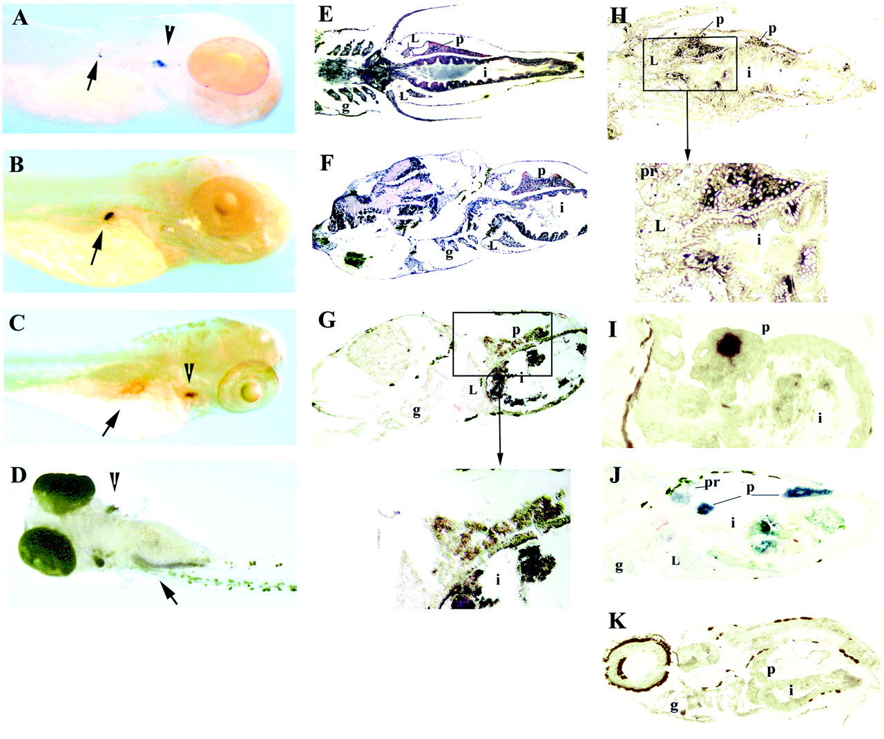
Fig. 3 Expression of Igμ and rag1 during development. Whole-mount in situ hybridization of 4-day fish with rag1 (A) and insulin (B) probes. In addition to strong staining with the rag1 probe in the thymus (A, arrowhead), weak staining can be detected in the right dorsal aspect of the abdominal cavity (A, arrow), where the developing pancreas is located; the location of staining with the insulin probe (B, arrow) appears to coincide with that by the rag1 probe. (C) Whole-mount in situ hybridization of 5-day fish with the rag1 probe. The arrowhead points to the thymus and the arrow to the pancreas; the central unstained area within the pancreas corresponds to an islet of Langerhans. (D) Whole-mount in situ hybridization of 8-day fish with rag1 probe, ventral view. Staining is seen in thymus (arrowhead) and pancreas (arrow); the pancreas is now elongated. Horizontal (E) and sagittal (F) paraffin sections through a 10-day-old fish stained with hematoxylin/eosin. The intestine at this age is a straight tube; the pancreas extends along the intestine. In situ hybridization of sagittal (G) or horizontal (H and I) cryosections of 10-day fish with Igμ (G), rag1 (H), or insulin (I) probes. Staining with rag1 and Igμ probes is seen in the pancreas surrounding an islet of Langerhans; with the rag1 probe, a few intraepithelial cells within the intestine also are stained. Intense staining of an islet is seen with the insulin probe. (J) In situ hybridization of a sagittal cryosection of 19-day fish with Igμ probe; most staining is within the pancreas, but some is also seen in the pronephros. (K) In situ hybridization of sagittal cryosection with Igμ sense probe; the brown color is pigment. L, liver; g, gills; i, intestine; p, pancreas; pr, pronephros. The fish in A is albino; those in B-D are wild-type treated with phenylthiourea and the rest are untreated wild-type. The dark material within the intestine in G-J is remnants of food.