
Fig. 5
- ID
- ZDB-FIG-080529-82
- Publication
- Danilova et al., 2002 - B cells develop in the zebrafish pancreas
- Other Figures
- All Figure Page
- Back to All Figure Page
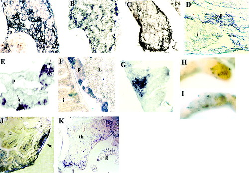
Expression of Igμ and rag1 in organs of adult (3 months old) zebrafish. In situ hybridization on transverse cryosections (A-C, E-G, J, and K) or sagittal paraffin section (D). Pronephros (A) Igμ and rag1 (B) probes. Stained cells form clusters. (C) Mesonephros, Igμ probe; stained cells are mostly in the periphery, with only a few internally. (D) Intestinal and pancreatic tissue, Igμ probe. Note intense staining in region of pancreas. (E) Higher magnification of pancreatic tissue, Igμ probe; note accumulation of stained cells near blood vessels (arrows). (F) Intestine and pancreas, rag1 probe. Stained cells within pancreas form clusters. Staining is also seen in the lamina propria. (G) Intestine, Igμ probe; note accumulation of Igμ+ cells in lamina propria. Whole-mount in situ hybridization of segment of intestine with Igμ (H) and rag1(I) probes; stained cells form patches. (J) Section across patch shown in H; Igμ+ cells cluster into a follicle-like aggregate (arrow). (K) Section through thymus (th), Igμ probe; note also staining in gills (g). |