
- Title
-
Trim45 is essential to the development of the diencephalon and eye in zebrafish embryos
- Authors
- Choe, S., Huh, T.L., Rhee, M.
- Source
- Full text @ Animal Cells Syst (Seoul)
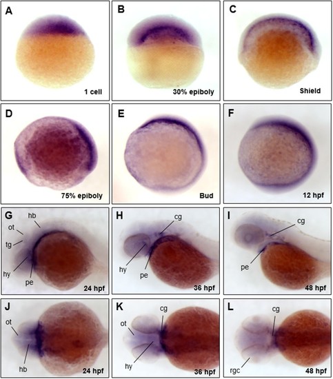
The spatio-temporal expression patterns of |
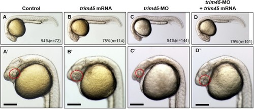
Phenotypes of the embryos from the overexpression or knock-down of |
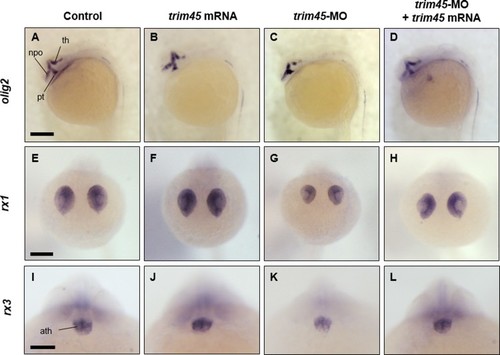
Spatio-temporal expression of |
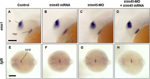
Spatio-temporal expression of |