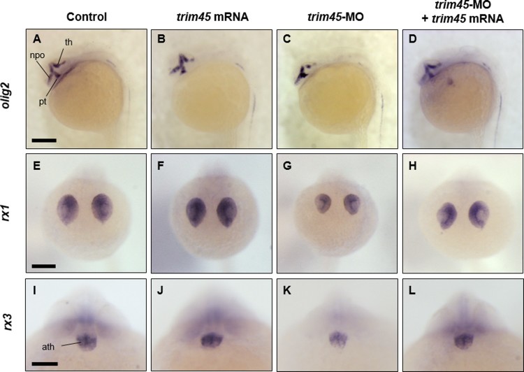
IMAGE

Figure 4.

ID
ZDB-IMAGE-200605-5
Source
Figures for Choe et al., 2020
Image
Figure Caption

Figure 4.

Spatio-temporal expression of olig2, rx1 and rx3, markers for diencephalon, retina, and optic primordia, in zebrafish embryos at 24 hpf. WISH was carried out with the following markers: olig2, rx1, and rx3 on each embryo (Figure 3 embryos were used). Overexpression of trim45 showed similar expression patterns with control for olig2 (A and B), rx1 (E and F), and rx3 (I and J). But, in knock-down, trim45 morphants showed compression of olig2 expression region and decrease in the size of thalamus (C). Also, knock-down of trim45 induced size reduction on eyes and decrease in transcript level of rx1 (G) and rx3 (K). Co-injection of trim45 mRNA with trim45-MO rescued the morphological phenotype of trim45 morphants (D, H and L). Lateral views – (A–D); anterior views – (E–L) th, thalamus; npo, neurosecretory preoptic area; pt, posterior tuberculum; ath, anterior hypothalamus. Scale bar – 200 μm (A–H), 100 μm (I–L).

Acknowledgments
This image is the copyrighted work of the attributed author or publisher, and ZFIN has permission only to display this image to its users. Additional permissions should be obtained from the applicable author or publisher of the image. Full text @ Animal Cells Syst (Seoul)