- Title
-
Transient and lineage-restricted requirement of Ebf3 for sternum ossification
- Authors
- Kuriki, M., Sato, F., Arai, H.N., Sogabe, M., Kaneko, M., Kiyonari, H., Kawakami, K., Yoshimoto, Y., Shukunami, C., Sehara-Fujisawa, A.
- Source
- Full text @ Development
|
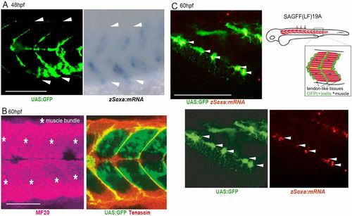
Identification of an enhancer trap line marking progenitors of tenocytes and interstitial cells in zebrafish embryos. (A) Expression of UAS:GFP in Gal4-UAS enhancer trap zebrafish line SAGFF(LF)19A and signals for scxa mRNA transcripts detected by whole-mount in situ hybridization (WISH) at 48 hpf. White arrowheads show segmental boundaries of somites. (B) Whole-mount immunostaining of 60 hpf SAGFF(LF)19A embryos for myogenic (magenta, MF20) and tenogenic (red, tenascin) markers together with UAS:GFP signals at 60 hpf. Asterisks mark muscle bundles. (C) Tenocyte progenitors marked with UAS:GFP and fluorescence in situ hybridization for scxa (red) at 60 hpf. Note that these cells extended long filopodia through the boundaries of skeletal muscle fibers. Arrowheads indicate UAS:GFP+ zScxa:mRNA+ cells. Schematic shows UAS:GFP+ cells in tendon and connective tissues in trunk muscles of SAGFF(LF) 19A line. Scale bars: 100 μm (A,B); 50 μm (C). |
|
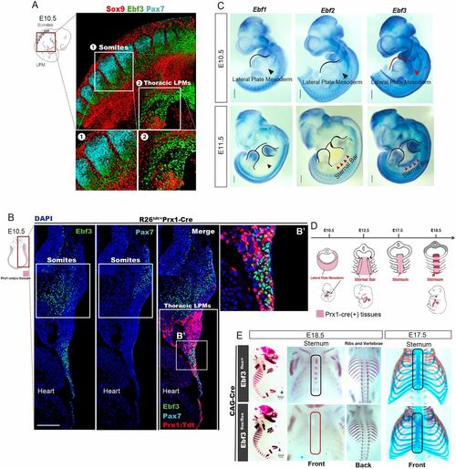
Essential roles of Ebf3 in sternum ossification. (A) Whole-mount immunostaining of E10.5 mouse embryo with antibodies against Ebf3 (green), Pax7 (cyan) and Sox9 (red). Ebf3 was expressed in the lateral plate mesoderm, as well as in syndetomes located between Pax7+ dermomyotomes and Sox9+ sclerotomes. Insets 1 and 2 show higher magnifications of the indicated regions. (B,B′) Ebf3 (green) was expressed in Prx1:Tdtomato+ cells (red) at the thoracic lateral plate mesoderm and in syndetomes adjacent to Pax7+ dermomyotomes (cyan) at E10.5. B′ shows an enlarged view of the boxed region. (C) WISH for Ebf1, Ebf2 and Ebf3 in E10.5 and E11.5 mouse embryos. Red arrowheads indicate that Ebf3, but not other Ebfs, was expressed in the thoracic lateral plate mesoderm tissue at E10.5 and show highest expression of Ebf3 in the sternal primordial bars at E11.5. (D) Schema of sternum development during murine embryogenesis. Red regions represent sternal bars at E12.5 and ossified zones of the sternum at E18.5. (E) Ossification and chondrogenesis analyses of Ebf3flox/flox CAG-Cre and Ebf3flox/+ CAG-Cre embryos at E18.5 and E17.5 based on skeletal preparations. Ebf3-KO mice exhibited defective sternum ossification without any defects in chondrogenesis. Scale bars: 100 μm (B); 1 mm (C); 2 mm (E). |
|
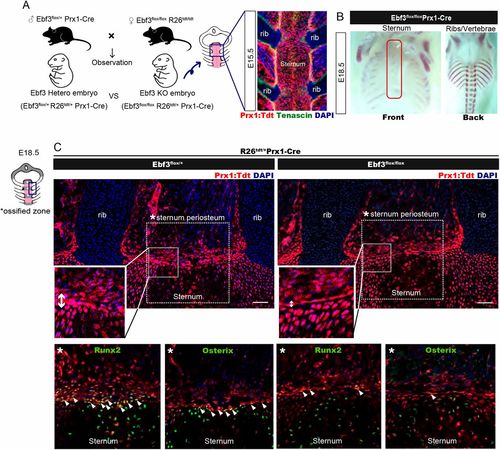
Ebf3 is required in lateral plate mesoderm-derived cells for sternum osteoblast differentiation. (A) Left: The generation of lateral plate mesoderm-specific Ebf3-KO mice embryos by crossing the Ebf3flox/+ Prx1-Cre line with Ebf3flox/flox Rosa26Tdtomato/Tdtomato mice. Right: The expression pattern of Prx1-Cre-activated Tdtomato in the sternum tissue and muscle connective tissues based on longitudinal sections of E15.5 embryos together with immunostaining for tenascin (green). (B) Ossification analysis based on Alizarin Red skeletal preparations of thoracic bones in Ebf3flox/flox Prx1-Cre embryos at E18.5. (C) Longitudinal sections of the sternum of Ebf3flox/flox Prx1-Cre and Ebf3flox/+ Prx1-Cre embryos at E18.5, immunostained for osteoblast (green, osterix) and pre-osteoblast (green, Runx2) markers. Upper panels show the distribution of lateral plate mesoderm-derived cells labeled with Prx1-Cre-activated TdTomato (red) in interstitial cells surrounding the sternum, which was decreased in Ebf3-KO embryos (n=7) compared with heterozygous mice (n=7). Lower-enlarged panels show much fewer osterix/Runx2-positive lateral plate mesoderm-derived cells in the periosteum of Ebf3-KO mice compared with heterozygous mice (white arrowheads). Scale bars: 100 μm. |
|
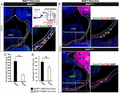
Decreased number of Runx2+ pre-osteoblasts in Ebf3-deficient thoracic lateral plate mesoderm at E12.5. (A) Transverse section of thoracic lateral plate mesoderm (as indicated in the schematic) at E12.5 immunostained with anti-Ebf3 and anti-Runx2 antibodies. Most but not all Ebf3-expressing cells were Runx2 positive. White and yellow arrowheads show Ebf3/Runx2 double-positive cells. (B) Decrease in the number of Prx1: Tdtomato+ Runx2+ cells in Ebf3flox/flox Prx1-Cre compared with that in heterozygous embryos (white arrowheads). Immunostained transverse sections of the thoracic lateral plate mesoderm at E12.5 are shown. A and upper panels of B show images of Ebf3/Runx2-double-positive cells and those of Prx1: Tdtomato+Runx2+ LPMs, respectively, prepared from a single section of a heterozygous embryo stained with antibodies against Ebf3, Runx2 and DAPI. Yellow arrowheads indicate Prx1: Tdtomato+Ebf3+Runx2+ LPMs. (C) The ratio of Runx2+ cells in Prx1: Tdtomato+ thoracic LPMs was lower in Ebf3flox/flox Prx1-Cre KO mice than in Ebf3 heterozygous mice at E12.5. Error bars represent s.e.m. **P<0.01 (n=4). (D) Ratio of Sox9+ cells in Prx1: Tdtomato+ thoracic LPMs at E12.5. Error bars represent s.e.m.; ns, non-significant (n=4). Scale bars: 100 μm |


