
|
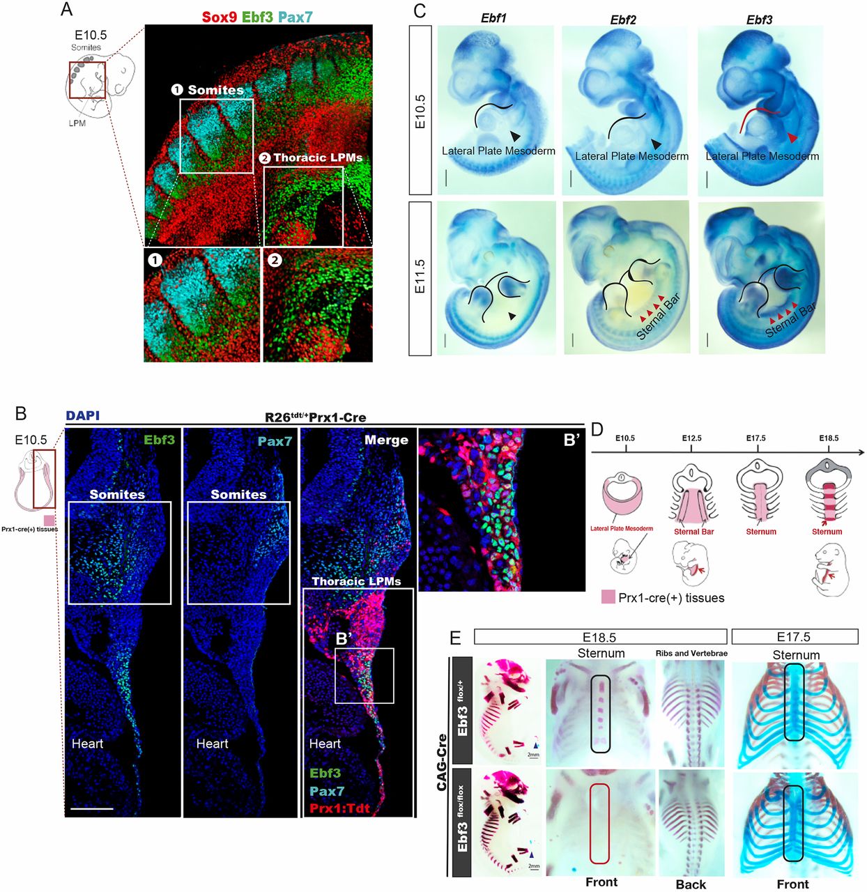
Fig. 2 Essential roles of Ebf3 in sternum ossification. (A) Whole-mount immunostaining of E10.5 mouse embryo with antibodies against Ebf3 (green), Pax7 (cyan) and Sox9 (red). Ebf3 was expressed in the lateral plate mesoderm, as well as in syndetomes located between Pax7+ dermomyotomes and Sox9+ sclerotomes. Insets 1 and 2 show higher magnifications of the indicated regions. (B,B′) Ebf3 (green) was expressed in Prx1:Tdtomato+ cells (red) at the thoracic lateral plate mesoderm and in syndetomes adjacent to Pax7+ dermomyotomes (cyan) at E10.5. B′ shows an enlarged view of the boxed region. (C) WISH for Ebf1, Ebf2 and Ebf3 in E10.5 and E11.5 mouse embryos. Red arrowheads indicate that Ebf3, but not other Ebfs, was expressed in the thoracic lateral plate mesoderm tissue at E10.5 and show highest expression of Ebf3 in the sternal primordial bars at E11.5. (D) Schema of sternum development during murine embryogenesis. Red regions represent sternal bars at E12.5 and ossified zones of the sternum at E18.5. (E) Ossification and chondrogenesis analyses of Ebf3flox/flox CAG-Cre and Ebf3flox/+ CAG-Cre embryos at E18.5 and E17.5 based on skeletal preparations. Ebf3-KO mice exhibited defective sternum ossification without any defects in chondrogenesis. Scale bars: 100 μm (B); 1 mm (C); 2 mm (E).