
|
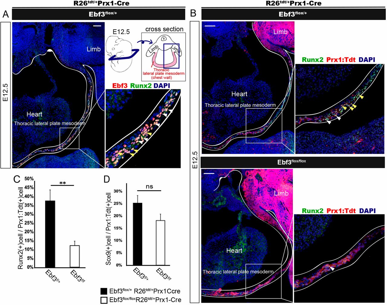
Fig. 4 Decreased number of Runx2+ pre-osteoblasts in Ebf3-deficient thoracic lateral plate mesoderm at E12.5. (A) Transverse section of thoracic lateral plate mesoderm (as indicated in the schematic) at E12.5 immunostained with anti-Ebf3 and anti-Runx2 antibodies. Most but not all Ebf3-expressing cells were Runx2 positive. White and yellow arrowheads show Ebf3/Runx2 double-positive cells. (B) Decrease in the number of Prx1: Tdtomato+ Runx2+ cells in Ebf3flox/flox Prx1-Cre compared with that in heterozygous embryos (white arrowheads). Immunostained transverse sections of the thoracic lateral plate mesoderm at E12.5 are shown. A and upper panels of B show images of Ebf3/Runx2-double-positive cells and those of Prx1: Tdtomato+Runx2+ LPMs, respectively, prepared from a single section of a heterozygous embryo stained with antibodies against Ebf3, Runx2 and DAPI. Yellow arrowheads indicate Prx1: Tdtomato+Ebf3+Runx2+ LPMs. (C) The ratio of Runx2+ cells in Prx1: Tdtomato+ thoracic LPMs was lower in Ebf3flox/flox Prx1-Cre KO mice than in Ebf3 heterozygous mice at E12.5. Error bars represent s.e.m. **P<0.01 (n=4). (D) Ratio of Sox9+ cells in Prx1: Tdtomato+ thoracic LPMs at E12.5. Error bars represent s.e.m.; ns, non-significant (n=4). Scale bars: 100 μm