FIGURE SUMMARY
Title

Zebrafish Avatars towards Personalized Medicine-A Comparative Review between Avatar Models

Authors
Costa, B., Estrada, M.F., Mendes, R.V., Fior, R.
Source
Full text @ Cells

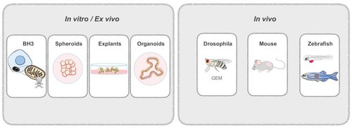
Patient-derived Avatars. Patient-derived cells are used to generate in vitro and in vivo Avatars. In vitro models include spheroids from dissociated tissue; explants, that are not dissociated and retain the original tissue architecture; and organoids, derived from adult stem cells. In vivo models include genetically engineered drosophila flies that mimic patient mutations; and patient-derived mouse or zebrafish xenografts. Zebrafish can be used at the larval or adult stage.

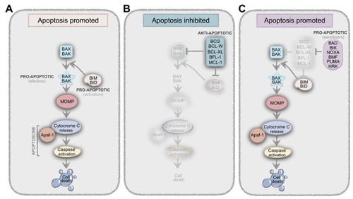
Balance between pro- and anti-apoptotic proteins result in life-death decisions. (A) BIM and BID (activators) are known as the BH3-only proteins whose function is to induce the conformational changes of BAX and BAK (effectors) to produce the Mitochondrial Outer Membrane Permeabilization (MOMP), resulting in apoptosis. (B) Anti-apoptotic proteins like BCL-2, BCL-W, BCL-XL, BFL-1, and MCL-1, can bind to BH3-only proteins to prevent their interaction with BAX and BAK, thus preventing apoptosis. (C) Another class of proteins, which are pro-apoptotic or sensitizers (BAD, BIK, NOXA, HRK, BMF, and PUMA) cannot directly bind to BAX and BAK, but instead bind to anti-apoptotic proteins, preventing the interaction between these and the activators.

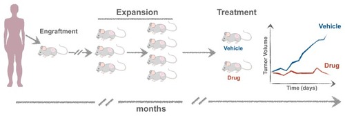
Experimental setup for generating mouse Patient-Derived Xenografts (mPDXs). The tumor is minced and transplanted either orthotopically or subcutaneously, embedded in matrigel. When the tumor reaches ~1 cm in diameter, it is excised and propagated into more mice (F2, F3) to obtain cohorts of Avatars where different therapies can be tested.

(A) Experimental setup for generating zebrafish xenografts. Cells derived from in vitro culture or primary human cells are labelled and microinjected in the PVS of 2dpf larvae. One day after injection, larvae are screened for successful injection and distributed in groups for testing chemo-, radio- and/or biological therapies. Three days after treatment, larvae (B) are fixed and processed for immunofluorescence for analysis of proliferation (C), cell death (D), angiogenesis (E), and metastatic potential (F,G). dpi: days post injection.

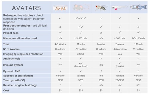
Comparison between patient-derived Avatar models.

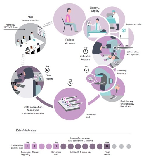
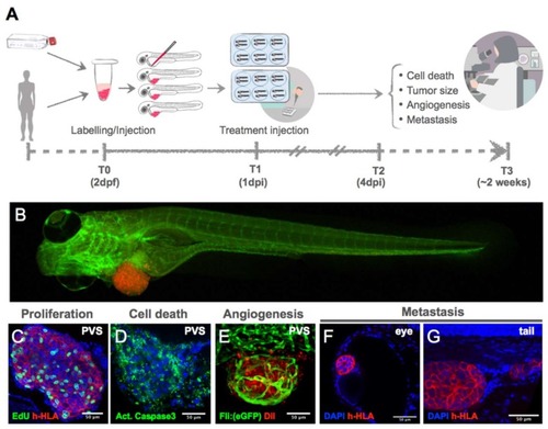
Workflow of zebrafish Avatars in the context of personalized medicine.

Acknowledgments
This image is the copyrighted work of the attributed author or publisher, and ZFIN has permission only to display this image to its users. Additional permissions should be obtained from the applicable author or publisher of the image. Full text @ Cells