
Image
|
Figure Caption
Figure 3
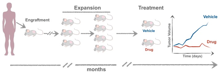
Experimental setup for generating mouse Patient-Derived Xenografts (mPDXs). The tumor is minced and transplanted either orthotopically or subcutaneously, embedded in matrigel. When the tumor reaches ~1 cm in diameter, it is excised and propagated into more mice (F2, F3) to obtain cohorts of Avatars where different therapies can be tested.
Acknowledgments
This image is the copyrighted work of the attributed author or publisher, and
ZFIN has permission only to display this image to its users.
Additional permissions should be obtained from the applicable author or publisher of the image.
Full text @ Cells