- Title
-
Midline crossing is not required for subsequent pathfinding decisions in commissural neurons
- Authors
- Bonner, J., Letko, M., Nikolaus, O.B., Krug, L., Cooper, A., Chadwick, B., Conklin, P., Lim, A., Chien, C.B., and Dorsky, R.I.
- Source
- Full text @ Neural Dev.
|
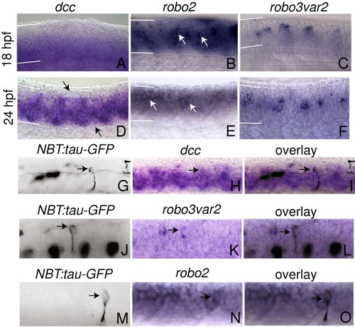
Expression of dcc, robo2 and robo3var2 in the zebrafish spinal cord. (A-C) Expression at 18 hpf. Adcc is expressed in the ventral half of the spinal cord. Brobo2 is expressed throughout the spinal cord and is observed in numerous postmitotic neurons (arrows). Crobo3var2 expression is restricted to postmitotic neurons at 18 hpf. D-F Expression at 24 hpf. Ddcc expression has expanded to include almost the entire spinal cord; however, expression is weaker in segmentally repeated regions in dorsal and ventral spinal cord, as indicated by arrows. Erobo2 is expressed in postmitotic neurons (arrows). F At 24 hpf, robo3var2 is expressed in postmitotic neurons. G-O Pseudocolored reflected light image of anti-GFP immunofluorescence in NBT:tau-GFP embryos (G, J, M) and transmitted light images of the same embryos that have undergone in situ hybridization for dcc, robo3var2, and robo2 (h, k, n) at 24 hpf. (i, l, o) exhibit overlain images of (G and H), (J and K), and (M and N), respectively. CoPA neurons are indicated by arrows. In all images, dorsal is up, anterior to the left. White lines indicate the dorsal and ventral boundaries of the spinal cord. |
|
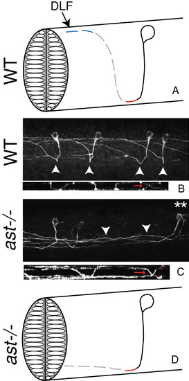
robo2 is required for escaping the midline and dorsal growth after crossing the midline. A Schematic representation of one CoPA neuron. Solid black line indicates the ipsilateral ventral projection, red line indicates the midline crossing of the commissure, the dotted gray line indicates dorsal pathfinding after crossing the midline, and dotted blue line indicates anterior pathfinding after crossing the midline. CoPA axons join the dorsal longitudinal fasciculus (DLF) to ascend. CoPA axons extend ventrally at 17 hpf (solid line), cross the midline at 18 hpf, extend dorsally at 19 hpf, and grow toward the head at 21 hpf. Timeline adapted from Kuwada et al., 1990. B Confocal micrograph of 3A10 immunofluorescence in the spinal cord illustrating wild-type CoPA pathfinding in multiple segments, lateral view. Arrowheads indicate midline crossing or commissures. In smaller image below, a dorsal view of the same spinal cord indicates midline crossing (red arrows) C In astti272z embryos, CoPA axons cross the midline, but remain ventral for several segments while ascending. Asterisks indicate affected CoPA cell bodies, and arrowheads mark axons that fail to extend dorsally after crossing the midline. In the smaller image below, a dorsal view of the same spinal cord indicates midline crossing (red arrows) of affected CoPA axons (cell bodies indicated by asterisks). D Summary diagram of astti272z phenotype. In all images dorsal is up, anterior to the left. |
|
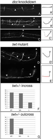
Crossing is not required for reception of anterior or dorsal guidance cues. A-Cdcc knockdown embryos demonstrating phenotypes consistent with loss of reception of attractive ventral cues, including anterior growth with a failure to grow ventrally (a′-c′) Summary diagrams illustrating affected axons. AAsterisk indicates affected CoPA, while arrowhead points to affected CoPA axon. In (B), an ipsilateral projection that grows dorsally and anteriorly without crossing. Asterisk and arrowheads indicate affected cell body and axons, respectively. C A ventral extension (arrowhead) from an affected CoPA (asterisk) grows at an oblique angle to the ventral spinal cord (compare to wild-type pathfinding, Figure 2a-b). (D-E) robo3 (twttx209) embryos exhibit two distinct phenotypes. D′-E′ Summary diagrams illustrating affected axons. In (D), robo3 (twttx209) CoPA axons fail to enter the midline and ascend instead on the ipsilateral side of the spinal cord. Asterisk indicates affected CoPA neuron, arrowheads indicate ipsilateral projection in this image, in which only the ipsilateral spinal cord was imaged. In (E) robo3 (twttx209), CoPA axons enter the midline but remain for some distance while ascending before exiting the midline on the contralateral side (“midline ascending”). Asterisk denotes CoPA cell body, arrowheads indicate axons ascending within the midline. In all images, dorsal is up, anterior to the left. (F-G) Comparative phenotypes from offspring of heterozygous incross (F) versus outcross to wild type (G). Only in offspring of heterozygous incross is the ipsilateral (IL) CoPA phenotype observed. The midline ascending (MA) phenotype is present in both conditions. |
|
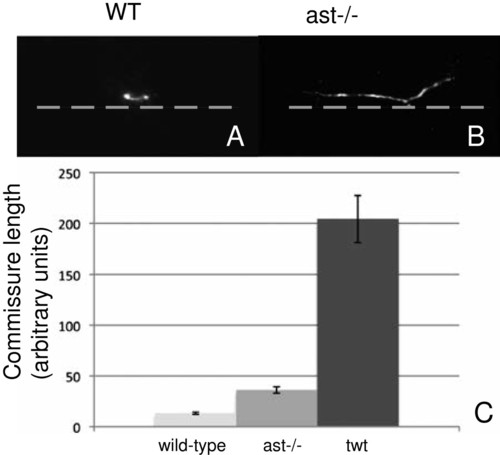
robo2 and robo3 are required for commissure formation. A-B Confocal microscopic images of WT and ast-/- commissures from individual CoPA axons (lateral view). astti272z CoPA axons ascend in the midline while WT CoPA axons cross immediately. Anterior is to the left, dorsal is up. Gray dotted line indicates ventral midline of spinal cord. C Measurements of axon length within the midline of the spinal cord, in the anteroposterior axis (arbitrary units) in wild-type, astti272z, and twt embryos. astti272z commissure length is 36 ± 3.2 (n = 7), which is 2.8 times that of wild type, which measures 13.3 ± 1.1 (n = 19) . In affected twttx209embryos, the average length is roughly 15 times that of WT (204 ± 22.9; n = 9). |


