
|
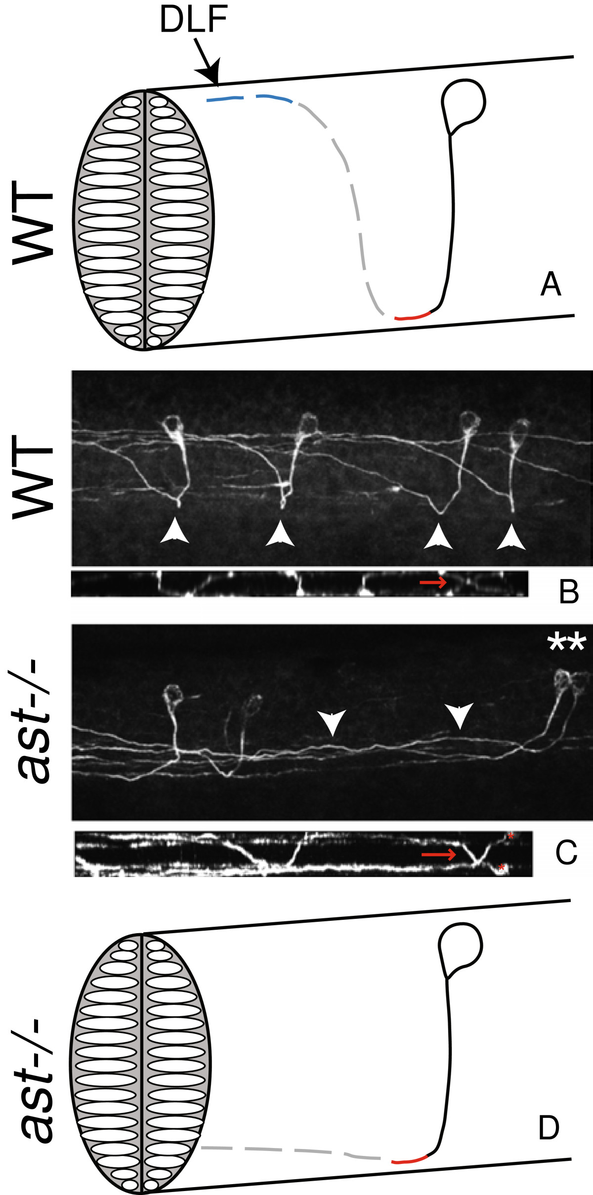
Fig. 2 robo2 is required for escaping the midline and dorsal growth after crossing the midline. A Schematic representation of one CoPA neuron. Solid black line indicates the ipsilateral ventral projection, red line indicates the midline crossing of the commissure, the dotted gray line indicates dorsal pathfinding after crossing the midline, and dotted blue line indicates anterior pathfinding after crossing the midline. CoPA axons join the dorsal longitudinal fasciculus (DLF) to ascend. CoPA axons extend ventrally at 17 hpf (solid line), cross the midline at 18 hpf, extend dorsally at 19 hpf, and grow toward the head at 21 hpf. Timeline adapted from Kuwada et al., 1990. B Confocal micrograph of 3A10 immunofluorescence in the spinal cord illustrating wild-type CoPA pathfinding in multiple segments, lateral view. Arrowheads indicate midline crossing or commissures. In smaller image below, a dorsal view of the same spinal cord indicates midline crossing (red arrows) C In astti272z embryos, CoPA axons cross the midline, but remain ventral for several segments while ascending. Asterisks indicate affected CoPA cell bodies, and arrowheads mark axons that fail to extend dorsally after crossing the midline. In the smaller image below, a dorsal view of the same spinal cord indicates midline crossing (red arrows) of affected CoPA axons (cell bodies indicated by asterisks). D Summary diagram of astti272z phenotype. In all images dorsal is up, anterior to the left.