
- Title
-
Multiple domains in the Crumbs Homolog 2a (Crb2a) protein are required for regulating rod photoreceptor size
- Authors
- Hsu, Y.C., and Jensen, A.M.
- Source
- Full text @ BMC Cell Biol.
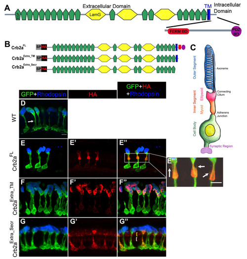
Localization of Crb2a extracellular domain-containing constructs in rod photoreceptors and transgenic rod morphology. Domain structure of zebrafish Crb2a protein. Crb2a is a single-pass transmembrane (blue) protein. The extracellular domain contains 19 EGF-like domains (green) and 3 laminin-like domains (yellow). The small intracellular domain contains two protein-protein interacting domains, the FERM-binding domain (FBD; red) and the PDZ-binding domain (PBD; purple). (B) Crb2a constructs containing the extracellular domain. All constructs have a signal peptide (SP) followed by an HA tag. Crb2aExtra_TM has the same sequence as Crb2aFL except the intracellular domain was deleted. Crb2aExtra_Secr has only the extracellular domain. (C) The rod photoreceptor has four morphologically and functionally distinct compartments: two basal compartments, the cell body (green) and synaptic region (purple), and two apical compartments, the inner segment (orange and pink) and outer segment (blue) filled with membranous discs packed with Rhodopsin. The inner segment is further subdivided into the myoid region (orange) and ellipsoid region (pink). The apical and basal compartments are separated by an adherens junction. (D-G″) Confocal z-projections of 6 d rods labeled with GFP (green), Rhodopsin (blue) and anti-HA (red) antibodies. (D) Wild-type rods. The adherens junction, the outer limiting membrane (OLM), is indicated by the white arrow. (E-E″′) Crb2aFL transgenic rods. Higher magnification of the inner segment showing fine processes (indicated by arrows) emerging from the inner segment (E″′). (F-F″) Crb2aExtra_TM transgenic rods. (G-G″) Crb2aExtra_Secr transgenic rods. Two rows of Crb2aExtra_Secr are indicated by yellow and white brackets, Scale bar, 5 μm (D-G″). EXPRESSION / LABELING:
|
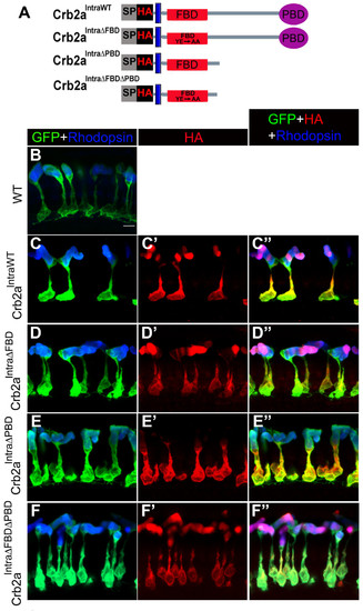
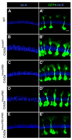
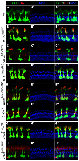
Localization of Crb2a constructs lacking the extracellular domain in rod photoreceptors and transgenic rod morphology. (A) Crb2a constructs lacking the extracellular domain. All constructs have a signal peptide (SP) followed by an HA tag. Crb2aIntraWT contains the transmembrane domain and intracellular sequence of Crb2a. Crb2aIntraΔFBD bears two mutations (Y10A and E16A) within the FERM-binding domain (FBD). Crb2aIntraΔPBD has the sequence following the FBD that contains the PDZ-binding domain (PBD) deleted. Crb2aIntraΔFBDΔPBD bears Y10A and E16A mutations within the FBD and the PBD the sequence following the FBD that contains the PDZ-binding domain (PBD) deleted. (B-F″) Confocal z-projections of 6 d rods labeled with GFP (green), Rhodopsin (blue) and anti-HA (red) antibodies. (B) Wild-type rods. (C-C″) Crb2aIntraWT transgenic rods. (D-D″) Crb2aIntraΔFBD transgenic rods. (E-E″) Crb2aIntraΔPBD transgenic rods. (F-F″) Crb2aIntraΔFBDΔPBD transgenic rods. Scale bar, 5 μm (B-F″). EXPRESSION / LABELING:
|
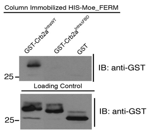
The FBD mutations Crb2a abolish binding to the FERM protein Moe. In vitro GST interaction between HIS-Moe_FERM immobilized on the column with GST-Crb2aIntraWT and GST-Crb2aIntraΔFBD. Loading control showing 1/40 input blotted and labeled with anti-GST. |
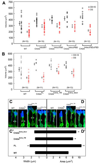
Measurement of rod size in Crb2a transgenics. (A) The average combined volume of rod cell body and inner segment (black circles) measured 332 ± 30 μm3 (wildtype), 372 ± 13.6 μm3 (Crb2aIntraWT), 394 ± 12.2 μm3 (Crb2aIntraΔFBD), 416 ± 28.9 μm3 (Crb2aIntraΔPBD), and 436 ± 22.8 μm3 (Crb2aIntraΔFBDΔPBD). The average volume of a rod outer segment (red circles) measured 190 ± 15.7 μm3 (wildtypes), 277 ± 15.4 μm3 (Crb2aIntraWT), 283 ± 20.6 μm3 (Crb2aIntraΔFBD), 278 ± 15.9 μm3 (Crb2aIntraΔPBD), and 212 ± 18.9 μm3 (Crb2aIntraΔFBDΔPBD). (B) The average combined volume of rod cell body and inner segment (black circles) measured 332 ± 30 μm3 (wildtype), 426 ± 17.5 μm3 (Crb2aFL), 401 ± 17.8 μm3 (Crb2aExtra_TM), and 405 ± 11 μm3 (Crb2aExtra_Secr). The average volume of a rod outer segment (red circles) measured 190 ± 15.7 μm3 (wildtype), 198 ± 13.8 μm3 (Crb2aFL), 316 ± 17.2 μm3 (Crb2aExtra_TM), and 268 ± 12.1 μm3 (Crb2aExtra_Secr). (C, C′) Width of the inner segment myoid region was marked as in (C) and the measurements are shown in (C′). The width of the myoid region is significantly larger in Crb2aFL rods (2.2 ± 0.1 μm) than wildtypes (1.5 ± 0.08 μm). Crb2aExtra_TM (1.5 ± 0.07 μm) and Crb2aExtra_Secr (1.6 ± 0.06 μm) are similar to wildtypes. (D, D′) Area of the myoid region was outlined as in (D) and the measurements are shown in (D′). The area of the myoid region is significantly larger in Crb2aFL (10.7 ± 0.5 μm2), Crb2aExtra_TM (8.1 ± 0.4 μm2), and Crb2aExtra_Secr (9.1 ± 0.5 μm2) compared to wildtypes (6.7 ± 0.2 μm2). Each circle represents a measurement of a single rod. Shown values are mean ± s.e.m, *, p < 0.005, student′s t test. |
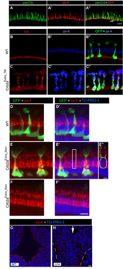
The zs-4 antibody recognizes the extracellular domain of Crb2a. (A-A″) Single confocal z-section of the photoreceptor layer in a 6 d wild-type retina labeled with rabbit panCrb antibodies (A) and zs-4 monoclonal (A′) and the two channels merged (A″). The arrow indicates where anti-panCrb and zs-4 labeling does not appear to fully overlap. (B-C″) Confocal z-projection of a 6 d retina labeled with anti-HA antibody (monoclonal IgG3; red) and zs-4 antibody (blue) and merged with anti-GFP (green). (B-B″) Wild-type retina. (C-C″) Crb2aExtra_TM transgenic retina. Endogenous Crb2a labeling is weak in comparison to Crb2aExtra_TM protein (C″). (D-F′) Confocal z-projection of the photoreceptor layer in a 6 d retina labeled with zs-4 antibody (red), rods express GFP (green). TO-PRO-3 (blue) labeling reveals the position of nuclei in the photoreceptor layer. (D-D′) Wild-type retina. (E-E″) Crb2aExtra_Secr transgenic retina. Single z-section from the white boxed area in E′, showing zs-4 labeling around the inner segment (indicated by yellow bracket and outlined in yellow) of a double-cone, the cell-body is circled in white (E″). (F-F′) Crb2aExtra_Secr transgenic retina where no rods are present. (G-H) Single z-section of the brain ventricular region of a 40 hour postfertilization embryo labeled with zs-4 antibody (red) showing ventricular surface labeling. Cell nuclei are labeled with TO-PRO-3 (blue). (G) Wild-type embryo. (H) ome (crb2a) mutant embryo. The midline (where the ventricles would normally form) is indicated by the white arrow, and eyes indicated by dashed white lines. Scale bars, 5 μm (A-A″′, D-F′) 10 μm (G, H). |
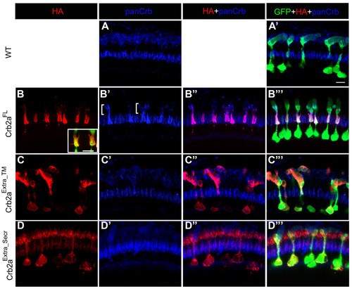
Effects of Crb2a extracellular domain-containing constructs on endogenous Crumbs proteins. (A-D″′) Confocal z-projections of 6 d rods labeled with GFP (green), panCrb (blue) and anti-HA (red) antibodies. (A, A′) Wild-type rods. (B-B″′) Crb2aFL transgenic rods. Inset in B shows fine processes emerging from the inner segment. Bracket in B′ indicates a region of strong panCrb labeling that is weakly labeled by anti-HA antibodies. (C-C″′) Crb2aExtra_TM transgenic rods. (D-D″′) Crb2aExtra_Secr transgenic rods. Scale bar, 5 μm. EXPRESSION / LABELING:
|
Effects of Crb2a constructs that lack the extracellular domain on endogenous Crb2a. (A-E′) Confocal z-projections of 6 d rods labeled with zs-4 (blue) and anti-GFP (green) antibodies. (A, A′) Wild-type rods. (B, B′) Crb2aIntraWT transgenic rods. (C, C′) Crb2aIntraΔFBD transgenic rods. (D, D′) Crb2aIntraΔPBD transgenic rods. (E, E′) Crb2aIntraΔFBDΔPBD transgenic rods. Scale bar, 5 μm. EXPRESSION / LABELING:
|
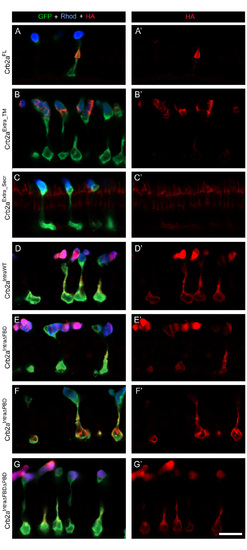
Single confocal z-sections of rods expressing Crb2a transgenes. (A, G′) Transgenic rods at 6 d labeled with GFP antibodies (green) and Rhodopsin antibodies (blue), and anti-HA antibodies (red). (A, A′) Crb2aFL transgenic rods. (B, B′) Crb2aExtra_TM transgenic rods. (C, C′) Crb2aExtra_Secr transgenic rods. (D, D′) Crb2aIntraWT transgenic rods. (E, E′) Crb2aIntraΔFBD transgenic rods. (F, F′) Crb2aIntraΔPBD transgenic rods. (G, G′) Crb2aIntraΔFBDΔPBD transgenic rods. Scale bar, 10 μm. EXPRESSION / LABELING:
|
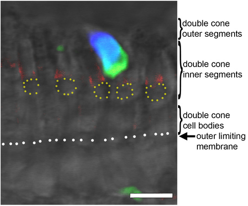
Crb2aExtra_Secr surrounds outer segments of cones. Single confocal z-section of a 6 d Crb2aExtra_Secr transgenic retina labeled with zs-4 antibodies (red), rhodopsin antibodies (blue) and anti-GFP antibodies (green) merged with the DIC-like image. Dotted yellow circles indicate the lipid droplet in double-cone inner segments, dotted white line indicates the outer limiting membrane. Scale bar, 5 μm. EXPRESSION / LABELING:
|
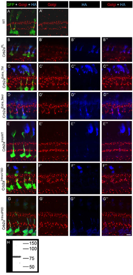
The Golgi apparatus, labeled by antibodies to Golgin subfamily A member 5 (GOLGA5), in 6 d wild-type and Crb2a transgenics. (A-G″′) Confocal z-projection of GFP-expressing rods in sections of 6 d retinas labeled with anti-GOLGA5 antibodies (red) and anti-HA (blue). (A, A′) Wild-type rods. (B-B″′) Crb2aFL transgenic rods. (C-C″′) Crb2aExtra_TM transgenic rods. (D-D″′) Crb2aExtra_Secr transgenic rods. (E-E″′) Crb2aIntraWT transgenic rods. (F-F″′) Crb2aIntraΔFBD transgenic rods. (G-G″′) Crb2aIntraΔPBD transgenic rods. (H) Western blot of 5 d wild-type zebrafish labeled with anti-GOLGA5 antibodies reveals a single protein of the expected molecular weight. The western blot was performed as described in [8], anti-GOLGA5 (Sigma HPA000992) was used at 1:1000 and HRP-conjugated goat anti-rabbit was used at 1:30,000. Scale bar, 5 μm. EXPRESSION / LABELING:
|
Effects of Crb2a construct expression on Moe localization. (A-H″) Confocal z-projection of 6 d photoreceptor layer labeled with anti-GFP (green), anti-HA antibodies (red) and anti-Moe antibodies (blue). (A-A′) Wild-type rods. (B-B″) Crb2aIntraWT transgenic rods. (C-C″) Crb2aIntraΔFBD transgenic rods. (D-D″) Crb2aIntraΔPBD transgenic rods. (E-E″) Crb2aIntraΔFBDΔPBD transgenic rods. (F-F″) Crb2aFL transgenic rods. (G-G″) Crb2aExtra_TM transgenic rods. (H-H″) Crb2aExtra_Secr transgenic rods. Scale bars, 5 μm. EXPRESSION / LABELING:
|
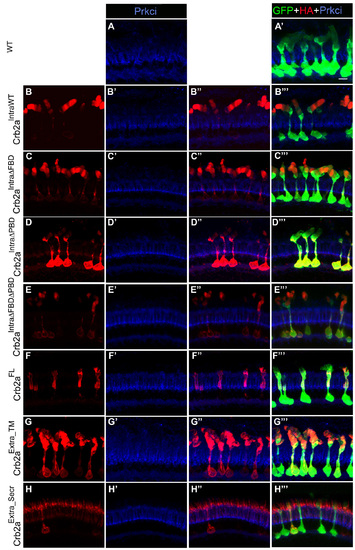
Effects of Crb2a construct expression on Prkci localization. (A-H″′) Confocal z-projection of 6 d photoreceptor layer labeled with anti-HA (red), anti-Prkci antibodies (blue) and anti-GFP labeling (green). (A-A″) Wild-type photoreceptor rods. (B-B″′) Crb2aIntraWT transgenic rods. (C-C″′) Crb2aIntraΔFBD transgenic rods. (D-D″′) Crb2aIntraΔPBD transgenic rods. (E-E″′) Crb2aIntraΔFBDΔPBD transgenic rods. (F-F″′) Crb2aFL transgenic rods. (G-G″′) Crb2aExtra_TM transgenic rods. (H-H″′) Crb2aExtra_Secr transgenic rods. Scale bars, 5 μm. |
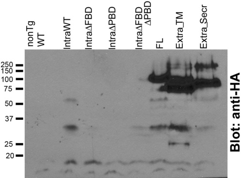
Western blot of 6 d wild-type (nonTg WT), Crb2aIntraWT, Crb2aIntraΔFBD, Crb2aIntraΔPBD, Crb2aIntraΔFBDΔPBD, Crb2aFL, Crb2aExtra_TM, Crb2aExtra_Secr transgenics probed with anti-HA antibodies. Western blotting was performed as previously described [8] and probed with anti-HA (clone 16B12) and HRP-conjugated goat anti-mouse. The predicted molecular weight of Crb2aIntraWT protein is 11 kD (retaining the signal peptide) but on western blot the major Crb2aIntraWT protein is ∼30kD, suggesting that it may be post-translationally modified or forms homoligomeres. Despite trying multiple gel and transfer conditions we were unable to detect Crb2aIntraΔFBD, Crb2aIntraΔPBD, Crb2aIntraΔFBDΔPBD proteins, which by immunohistochemistry are expressed at similar levels as Crb2aIntraWT. It is possible that Crb2aIntraΔFBD, which is be predicted to be about the same molecular weight as Crb2aIntraWT, is not post-translationally modified or does not dimerize and, thus, is too small, like Crb2aIntraΔPBD and Crb2aIntraΔFBDΔPBD with predicted molecular weights ∼8.8kD (with signal peptide) to be captured by Western blot analysis. |