
|
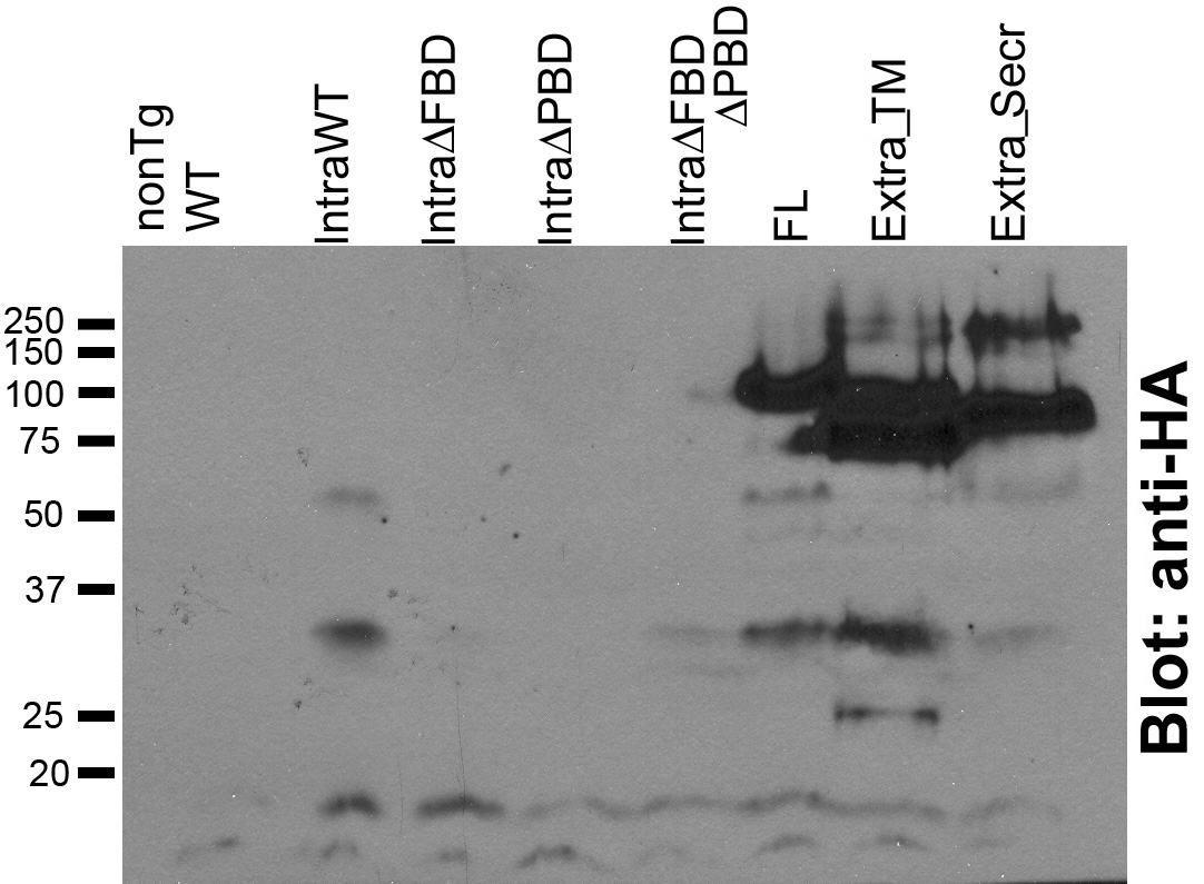
Fig. S6 Western blot of 6 d wild-type (nonTg WT), Crb2aIntraWT, Crb2aIntraΔFBD, Crb2aIntraΔPBD, Crb2aIntraΔFBDΔPBD, Crb2aFL, Crb2aExtra_TM, Crb2aExtra_Secr transgenics probed with anti-HA antibodies. Western blotting was performed as previously described [8] and probed with anti-HA (clone 16B12) and HRP-conjugated goat anti-mouse. The predicted molecular weight of Crb2aIntraWT protein is 11 kD (retaining the signal peptide) but on western blot the major Crb2aIntraWT protein is ∼30kD, suggesting that it may be post-translationally modified or forms homoligomeres. Despite trying multiple gel and transfer conditions we were unable to detect Crb2aIntraΔFBD, Crb2aIntraΔPBD, Crb2aIntraΔFBDΔPBD proteins, which by immunohistochemistry are expressed at similar levels as Crb2aIntraWT. It is possible that Crb2aIntraΔFBD, which is be predicted to be about the same molecular weight as Crb2aIntraWT, is not post-translationally modified or does not dimerize and, thus, is too small, like Crb2aIntraΔPBD and Crb2aIntraΔFBDΔPBD with predicted molecular weights ∼8.8kD (with signal peptide) to be captured by Western blot analysis.