
Image
|
Figure Caption
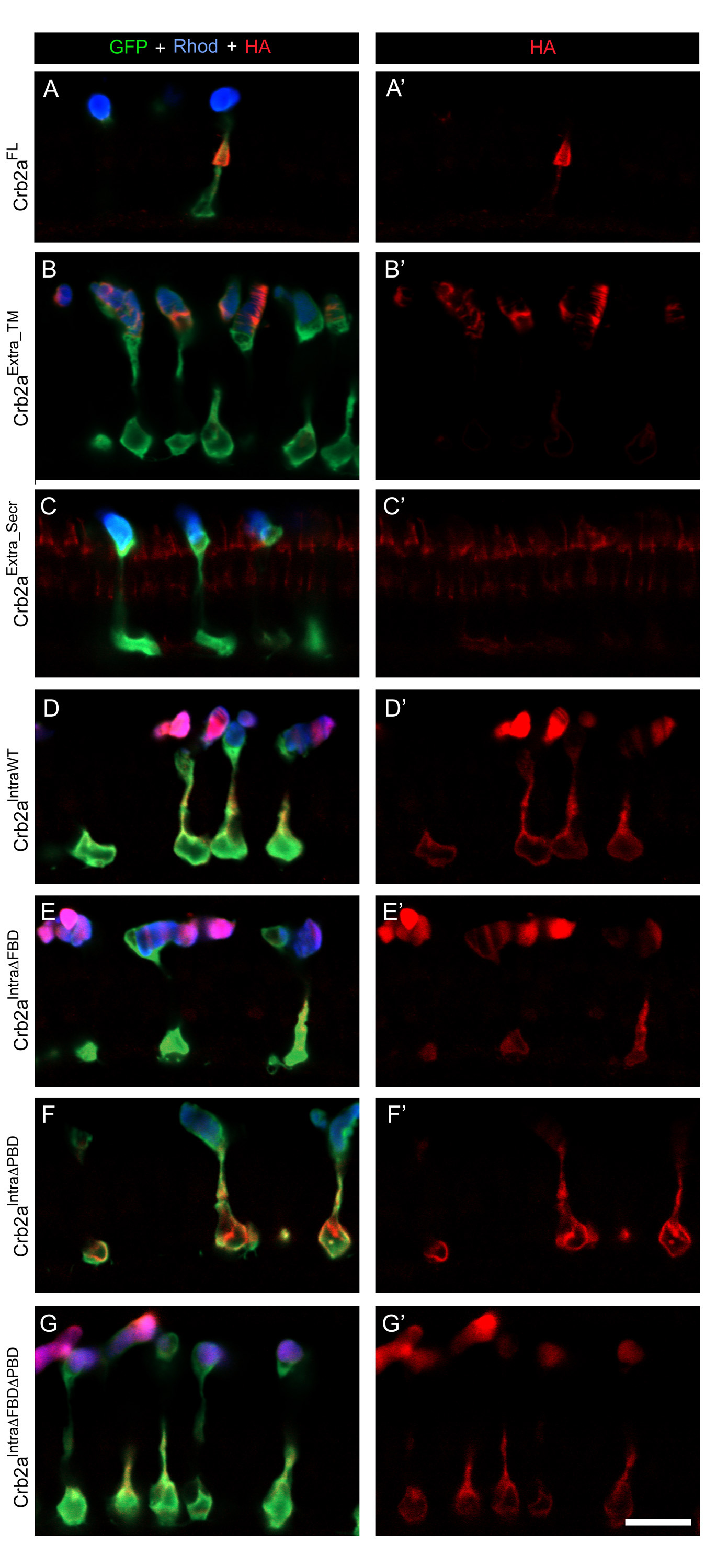
Fig. S1 Single confocal z-sections of rods expressing Crb2a transgenes. (A, G′) Transgenic rods at 6 d labeled with GFP antibodies (green) and Rhodopsin antibodies (blue), and anti-HA antibodies (red). (A, A′) Crb2aFL transgenic rods. (B, B′) Crb2aExtra_TM transgenic rods. (C, C′) Crb2aExtra_Secr transgenic rods. (D, D′) Crb2aIntraWT transgenic rods. (E, E′) Crb2aIntraΔFBD transgenic rods. (F, F′) Crb2aIntraΔPBD transgenic rods. (G, G′) Crb2aIntraΔFBDΔPBD transgenic rods. Scale bar, 10 μm.
Figure Data
Acknowledgments
This image is the copyrighted work of the attributed author or publisher, and
ZFIN has permission only to display this image to its users.
Additional permissions should be obtained from the applicable author or publisher of the image.
Open Access.
Full text @ BMC Cell Biol.