
- Title
-
Chromosomal position mediates spinal cord expression of a dbx1a enhancer
- Authors
- Gribble, S.L., Nikolaus, O.B., Carver, M.S., Hoshijima, K., and Dorsky, R.I.
- Source
- Full text @ Dev. Dyn.
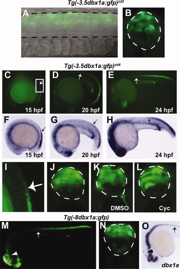
Upstream enhancer fragments of dbx1a drive reporter expression in a non-endogenous pattern. A: Lateral view of a live 24-hpf Tg(-3.5dbx1a:gfp)zd2 transgenic embryo. Spinal cord is outlined in dashed lines. B: Cross-section of a fixed embryo expressing the same transgene. In both views, GFP expression extends from the intermediate progenitor region to the dorsal limit of the spinal cord. C-E: Lateral views of live Tg(-3.5dbx1a:gfp)zd4 embryos. Arrows point to the anterior limit of transient spinal cord expression. F-H: In situ hybridization for gfp mRNA from the same insertion line. Arrows point to the anterior limit of transient spinal cord expression. At each stage, mRNA expression ceases slightly posterior to the limit of GFP protein. I: Enlarged image of the boxed region in C, showing loss of GFP expression anteriorly (arrow). J: Cross-section through the posterior spinal cord of a 24-hpf Tg(-3.5dbx1a:gfp)zd4 embryo. Reporter expression extends from the intermediate progenitor region to the dorsal limit of the spinal cord. K, L: Embryos treated with cyclopamine from 50% epiboly to 24 hpf show no ventral expansion of transgene expression compared with DMSO-treated controls. M: Lateral view of a live 24-hpf transgenic embryo expressing the dbx1a-8kb reporter from a Tol2 insertion. Arrow points to the anterior limit of transient spinal cord expression, and arrowhead marks tectal expression. N: Cross-section of a fixed embryo expressing the same transgene. O: Whole-mount in situ hybridization for dbx1a expression at 24 hpf, showing that endogenous expression is absent from the tectum (arrowhead) and present in the rostral spinal cord (arrow). |
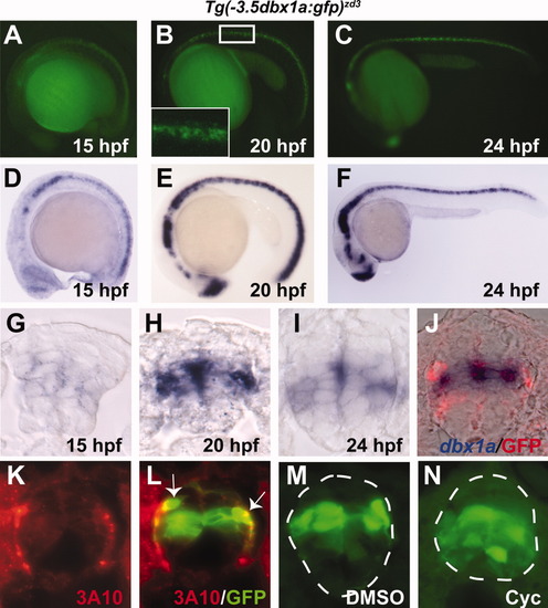
A single insertion line on chromosome 2 [Tg(-3.5dbx1a:gfp)zd3] expresses the dbx1a-3.5kb enhancer in an endogenous pattern. A-C: Lateral views of live Tg(-3.5dbx1a:gfp)zd3 embryos. Boxed region in B is enlarged in inset. D-F: In situ hybridization for gfp mRNA from the same insertion line. Starting at 15 hpf, GFP protein and mRNA are expressed throughout the anterior/posterior extent of the spinal cord. G-I: Cross-sections through the spinal cord of transgenic embryos, stained for gfp mRNA. J: Double staining for dbx1a mRNA (blue) and GFP protein (red). K, L: Staining with 3A10 antibody (red), which marks CoPA interneurons, and GFP (green). Arrows indicate double-labeled CoPA cells. M, N: Embryos treated with cyclopamine from 50% epiboly to 24 hpf show ventral expansion of transgene expression compared with DMSO-treated controls. |