
Fig. 3
- ID
- ZDB-FIG-091113-68
- Publication
- Gribble et al., 2009 - Chromosomal position mediates spinal cord expression of a dbx1a enhancer
- Other Figures
- All Figure Page
- Back to All Figure Page
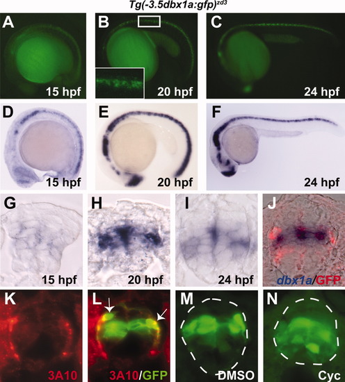
A single insertion line on chromosome 2 [Tg(-3.5dbx1a:gfp)zd3] expresses the dbx1a-3.5kb enhancer in an endogenous pattern. A-C: Lateral views of live Tg(-3.5dbx1a:gfp)zd3 embryos. Boxed region in B is enlarged in inset. D-F: In situ hybridization for gfp mRNA from the same insertion line. Starting at 15 hpf, GFP protein and mRNA are expressed throughout the anterior/posterior extent of the spinal cord. G-I: Cross-sections through the spinal cord of transgenic embryos, stained for gfp mRNA. J: Double staining for dbx1a mRNA (blue) and GFP protein (red). K, L: Staining with 3A10 antibody (red), which marks CoPA interneurons, and GFP (green). Arrows indicate double-labeled CoPA cells. M, N: Embryos treated with cyclopamine from 50% epiboly to 24 hpf show ventral expansion of transgene expression compared with DMSO-treated controls. |