- Title
-
Craniofacial skeletal defects of adult zebrafish Glypican 4 (knypek) mutants
- Authors
- LeClair, E.E., Mui, S.R., Huang, A., Topczewska, J.M., and Topczewski, J.
- Source
- Full text @ Dev. Dyn.
|
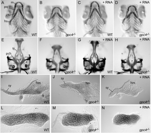
gpc4 mRNA fails to rescue craniofacial cartilage morphology of gpc4-/- (knypek) embryos. A-D: Ventral view of zebrafish pharyngeal arch cartilages stained with Alcian Blue at 7 days post-fertilization (dpf). Compared to wild-type siblings (A), the palatoquadrate, ceratohyal, and symplectic cartilages of gpc4-/- larvae were shorter and thicker (B). Wild type siblings injected with synthetic gpc4 mRNA developed normally (C). Injection of the same dose of synthetic mRNA fully rescued gastrulation defects of gpc4-/- embryos (Topczewski et al. [2001]), but the mutant cartilage phenotype was not suppressed (D). E-H: Ventral view of isolated larval neurocrania (7 dpf). Compared to wild types (E), the anterior ethmoid plate (ep) of gpc4-/- mutant larvae was shortened and the trabeculae (tr) were farther apart, causing an enlarged hypophyseal fenestra (hpf) (F). The posterior parachordal plate (pch) was relatively normal. Wild type siblings injected with synthetic gpc4 mRNA developed normal neurocrania (G), but in injected mutants the neurocranial defect was not suppressed (H). I-K: Flat-mounted hyosymplectic cartilages (7 dpf). Compared to wild types (I), gpc4-/- larvae showed a severely shortened symplectic region (sy, J). The hyomandibular region (hm) was less affected. This defect in gpc4-/- larvae was not suppressed by gpc4 mRNA injection (K). L-N: Flat-mounted ceratohyal cartilages (7 dpf). The ceratohyal of a wild type embryo (L) is much longer than that of a gpc4-/- embryo (M). The failure of ceratohyal cartilage elongation is not suppressed by injecting gpc4 mRNA into a gpc4-/- embryo (N). ch, ceratohyal; ep, ethmoid plate; hm, hyomandibular; hpf, hypopophyseal fenestra; hs, hyosymplectic; m, Meckel′s cartilage; pch, parachordal plate; pq, palatoquadrate; sy, symplectic; tr, trabeculae. PHENOTYPE:
|
|
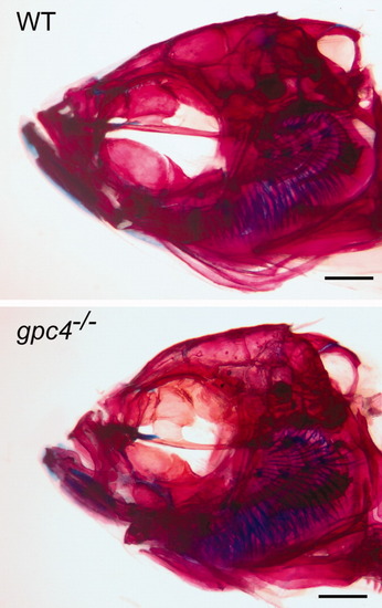
Cranial morphology of rescued gpc4-/- (knypek) mutants. Top: Wild type adult. Bottom: gpc4-/- mutant adult. Both fish are >1 year of age and are photographed at the same scale (scale bar = 1 mm). gpc4-/- fish are similar to comparable wild type fish in size and overall body proportions (data not shown) but have smaller heads, a domed skull, and shorter jawbones. |
|
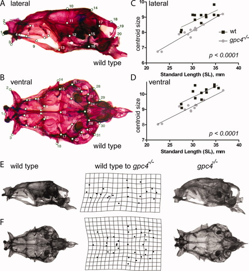
Size and shape comparison of wild type and gpc4-/- neurocraniaLocations of 20 lateral (A) and 33 ventral (B) skull landmarks used in the study. A complete description of each landmark is given in Supp. Table S1. In both views, a wild type skull is shown. C, D: Linear regression of lateral (C) and ventral (D) skull centroid size versus standard length for wild type (wt) and gpc4-/- mutant zebrafish. gpc4-/- neurocrania are significantly smaller than wild type neurocrania across a wide range of body sizes, as indicated by the significant downward shift in the gpc4-/- regression line (P < 0.0001 for differences in intercept). Lateral (E) and ventral (F) views of a wild type (left column) and gpc4-/- (right column) zebrafish skull. Center column: Deformation grid of neurocranial landmarks used in the study, derived from Procrustes superimposition of all wild type (n = 12) and gpc4-/- (n = 12) specimens. The grid indicates how the configuration of landmarks (A, B) must be altered to “convert” a wild type skull (left) into a mutant skull (right). Perfectly square cells indicate no difference in mean shape between mutants and wild types; deviations from squares indicate the regions of local shape difference between the two groups. |
|
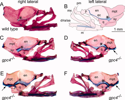
Loss of the symplectic bone in gpc4-/- mutant zebrafish. A: Right lateral facial bones of a wild type zebrafish, stained with Alcian Blue/Alizarin Red. Anterior is to the right. *, palatoquadrate cartilage. B: Mirror-image illustration of the facial bones and cartilages shown in A. Anterior is to the left. C, D: Unilateral loss of the symplectic in a gpc4-/- adult. The right side facial bones are normal (C); however, the left side of the same individual (D) lacks the symplectic (sy) ventral to the metapterygoid bone (mpt). In the absence of the symplectic, the metapterygoid and quadrate bones are attached by an ectopic band of cartilage (black arrow, D-F). A stub of cartilage fills the notch in the quadrate where the symplectic normally lies (white arrowhead, D-F). Note that relative to wild type (A), the palatoquadrate cartilage in gpc4-/- individuals appears broader and more deeply stained with Alcian Blue (asterisk, A-F). E, F: Bilateral loss of the symplectic in a second gpc4-/- adult. The morphology of both sides is similar to D. d/ra/aa, dentary/retroarticular/angular; ec, ectopterygoid; en, entopterygoid; m, Meckel′s cartilage; mx, maxilla; mpt, metapterygoid; p, palatine; pm, premaxilla; q, quadrate; sy, symplectic. All images are at the same magnification. Scale bar = 1 mm. |
|
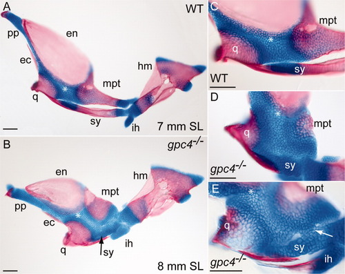
Intermediate stages of symplectic bone development in gpc4-/- larvae. A: Flat-mounted facial bones and cartilages from a 7-mm wild type larva (SL = standard length). Anterior is to the left. At this stage, the symplectic (sy) is a separate bone with two cartilaginous ends. B: The corresponding region of an 8-mm gpc4-/- larva. The reduced symplectic cartilage (sy, black arrow) has not ossified and is fused to the adjacent palatoquadrate. All other ossification centers are present. C: Magnification of the wild type embryo in A. D: Magnified palatoquadrate cartilage in a second gpc4-/- larva. The reduced symplectic (sy) is a ball of cartilage broadly connected with the palatoquadrate cartilage (*). E: Symplectic region of a third gpc4-/- larva. A cluster of chondrocytes (arrow) forms a bridge between the reduced symplectic and the palatoquadrate. The quadrate is only partially mineralized. Abbreviations are as in Figures 1 and 4. ih, interhyal; pp, pterygoid process. Scale bar = 100 μm. |
|
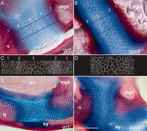
Adult gpc4-/- zebrafish lack elongated chondrocytes in the adult and juvenile palatoquadrate cartilage. A, B: Magnification of the adult cartilage band connecting the quadrate and metapterygoid bones. The wild type cartilage (A) has three regions: (1) bone overlapping with adjacent cartilage, (2) elongated chondrocytes parallel to the bone border, and (3) a central region of larger, rounded chondrocytes. In gpc4-/- adults, all three regions are modified (B). There is reduced overlap between bone and cartilage and a thickened bone border (region 1), no strongly elongated chondrocytes (region 2), and darker Alcian Blue staining overall (2 and 3). C, D: Outlines of cells traced from the regions between the dotted lines in A and B, respectively. Note the elongated cell boundaries in wild type region 2, which are absent in gpc4-/- mutants. E, F: The adult defect of chondrocyte organization originates in the early larval palatoquadrate (∼8 mm SL). The elongated cartilage cells present in wild type larvae (E) are rounded in gpc4-/- mutants (F). en, endopterygoid; mpt, metapterygoid; q, quadrate; sy, symplectic. Scale bar = 100 μm. |
|
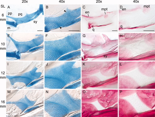
Developmental series of zebrafish palatoquadrate ossification.Flat-mounted views of larval zebrafish palatoquadrates. Anterior is to the left. For description, see text. SL, standard length of the fish (in mm). A-D: 6 mm SL. E-H: 10 mm SL. I-L: 12 mm SL. M-P: 16 mm SL. Stained specimens were cleared in 50% glycerol, flattened under a coverslip, and imaged with Nomarski optics. The images in the second and fourth columns (40x) are optical magnifications of the specimens in the first and third columns (20x). All images in a column are shown at the same magnification. Abbreviations are as in Figure 5. Arrowheads point to the arcs of elongated chondrocytes. All scale bars = 100 μm. |
|
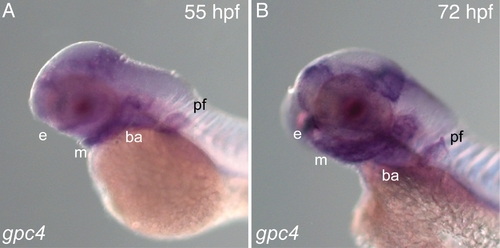
Expression of gpc4 during craniofacial cartilage morphogenesis. At 55 hpf (A) and 72 hpf (B), glypican 4 is strongly expressed in the regions where the neurocranium and pharyngeal cartilage elements are formed. e, ethmoid plate; m, Meckel’s cartilage; ba, branchial arches; pf, pectoral fin. |
|
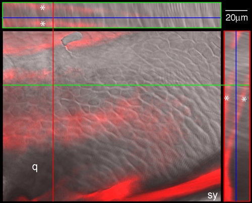
Perichondrial ossification of the zebrafish palatoquadrate. The central panel shows a magnified view of the palatoquadrate cartilage in a wild type zebrafish larva. Anterior is to the left. The tissue is stained with Alizarin Red to view early bone deposition. An orthogonal reconstruction of a z-stack through the crosshairs shows two layers of a bone matrix (double asterisks) enveloping a sheet of chondrocytes (visualized by Nomarski optics). q, region of the future quadrate bone, which is not yet undergoing endochondral replacement; sy, early ossification of the symplectic. |

