
|
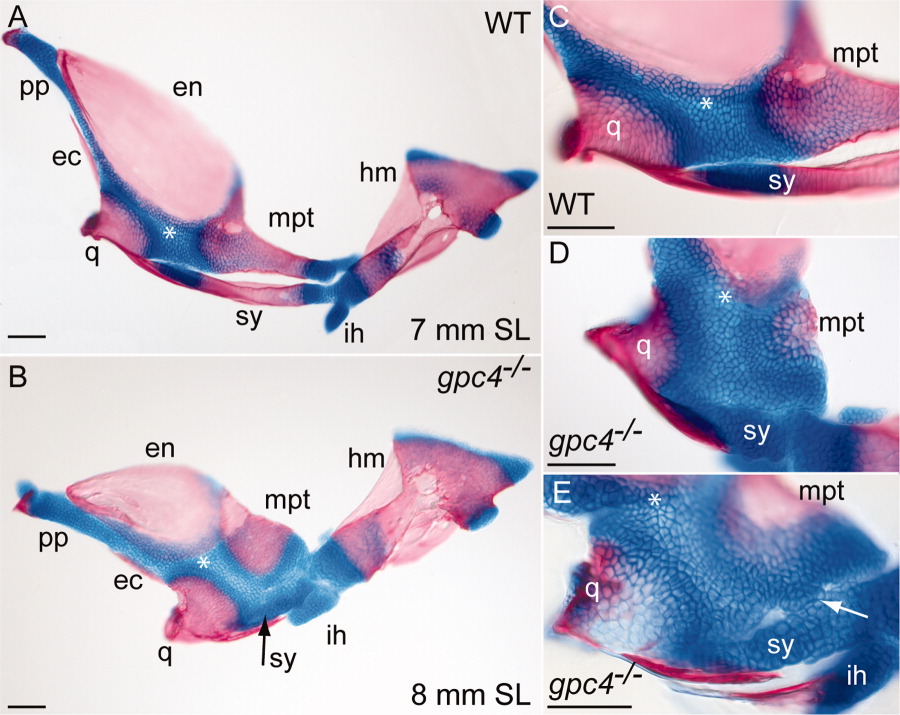
Fig. 5 Intermediate stages of symplectic bone development in gpc4-/- larvae. A: Flat-mounted facial bones and cartilages from a 7-mm wild type larva (SL = standard length). Anterior is to the left. At this stage, the symplectic (sy) is a separate bone with two cartilaginous ends. B: The corresponding region of an 8-mm gpc4-/- larva. The reduced symplectic cartilage (sy, black arrow) has not ossified and is fused to the adjacent palatoquadrate. All other ossification centers are present. C: Magnification of the wild type embryo in A. D: Magnified palatoquadrate cartilage in a second gpc4-/- larva. The reduced symplectic (sy) is a ball of cartilage broadly connected with the palatoquadrate cartilage (*). E: Symplectic region of a third gpc4-/- larva. A cluster of chondrocytes (arrow) forms a bridge between the reduced symplectic and the palatoquadrate. The quadrate is only partially mineralized. Abbreviations are as in Figures 1 and 4. ih, interhyal; pp, pterygoid process. Scale bar = 100 μm.