
FIGURE
Fig. S2
Fig. S2
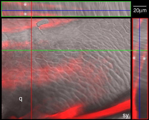
Perichondrial ossification of the zebrafish palatoquadrate. The central panel shows a magnified view of the palatoquadrate cartilage in a wild type zebrafish larva. Anterior is to the left. The tissue is stained with Alizarin Red to view early bone deposition. An orthogonal reconstruction of a z-stack through the crosshairs shows two layers of a bone matrix (double asterisks) enveloping a sheet of chondrocytes (visualized by Nomarski optics). q, region of the future quadrate bone, which is not yet undergoing endochondral replacement; sy, early ossification of the symplectic. |
Expression Data
Expression Detail
Antibody Labeling
Phenotype Data
Phenotype Detail
Acknowledgments
This image is the copyrighted work of the attributed author or publisher, and
ZFIN has permission only to display this image to its users.
Additional permissions should be obtained from the applicable author or publisher of the image.
Full text @ Dev. Dyn.