
|
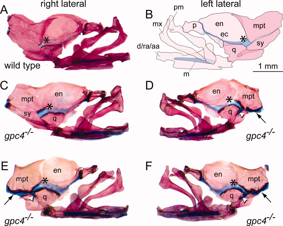
Fig. 4 Loss of the symplectic bone in gpc4-/- mutant zebrafish. A: Right lateral facial bones of a wild type zebrafish, stained with Alcian Blue/Alizarin Red. Anterior is to the right. *, palatoquadrate cartilage. B: Mirror-image illustration of the facial bones and cartilages shown in A. Anterior is to the left. C, D: Unilateral loss of the symplectic in a gpc4-/- adult. The right side facial bones are normal (C); however, the left side of the same individual (D) lacks the symplectic (sy) ventral to the metapterygoid bone (mpt). In the absence of the symplectic, the metapterygoid and quadrate bones are attached by an ectopic band of cartilage (black arrow, D-F). A stub of cartilage fills the notch in the quadrate where the symplectic normally lies (white arrowhead, D-F). Note that relative to wild type (A), the palatoquadrate cartilage in gpc4-/- individuals appears broader and more deeply stained with Alcian Blue (asterisk, A-F). E, F: Bilateral loss of the symplectic in a second gpc4-/- adult. The morphology of both sides is similar to D. d/ra/aa, dentary/retroarticular/angular; ec, ectopterygoid; en, entopterygoid; m, Meckel′s cartilage; mx, maxilla; mpt, metapterygoid; p, palatine; pm, premaxilla; q, quadrate; sy, symplectic. All images are at the same magnification. Scale bar = 1 mm.