
- Title
-
Transcriptional control of Rohon-Beard sensory neuron development at the neural plate border
- Authors
- Rossi, C.C., Kaji, T., and Artinger, K.B.
- Source
- Full text @ Dev. Dyn.
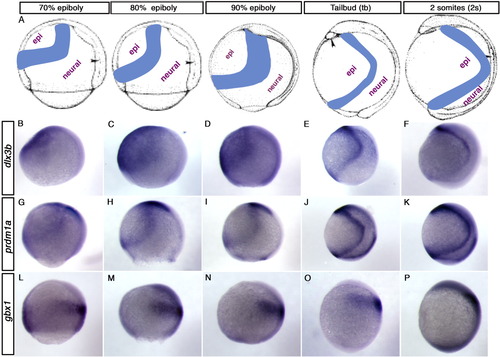
Detailed analysis of gene expression at early NPB stages. A-P: Lateral views of wild type embryos with dorsal toward the right and anterior toward the top. A: Schematic of the approximate location of the NPB, between dorsal neural ectoderm and ventral epidermal ectoderm, in gastrula stage zebrafish embryos. B-F: dlx3b expression from 70% epiboly through 2 somites. dlx3b transcripts are found throughout the epidermal ectoderm at 70% epiboly (B), 80% epiboly (C), and 90% epiboly (D). At tail bud, the expression domain narrows to a more discrete domain near the NPB (E), and at 2 somites the posterior portion of this domain is mostly gone (F). G-K: prdm1a expression. prdm1a transcripts are found at the NPB at each of these stages, but the domain narrows and moves toward the dorsal midline over time. Strong expression is also evident in the prechordal plate at the anterior tip of the embryo. L-P: gbx1 is expressed in dorsal neural tissue, and its domain shifts toward the future anterior as development proceeds. A was modified from Kimmel et al. ([1995]), with permission from the publisher. |
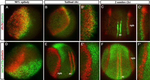
Overlap of the prdm1a expression domain with dlx3b is greater than with neural markers sox3 and sox19a. Confocal micrographs of wild type embryos mounted to best observe potential overlap of expression at the NPB; (A-B′,D) mounted laterally with dorsal toward the right, (C-C′,F-F′) mounted dorsally with anterior toward the top, and (E-E′) mounted dorsolaterally with anterior toward the top. A-C′: prdm1a in green, dlx3b in red, overlap in yellow. D: prdm1a in green, sox3 in red, overlap in yellow. E-F′: prdm1a in red, sox19a in green. The prdm1a domain overlaps extensively with dlx3b at 90% epiboly (yellow in A), with less overlap at tail bud (B,B′), and very little overlap by 2 somites (C,C′). D: A small amount of overlap of prdm1a and sox3 is observed at 90% epiboly (yellow). Transcripts for sox19a (green) and prdm1a (red) abut one another, but do not overlap with each other, at tail bud (E-E′) and 2 somites (F-F′). This is especially evident at 20x (E′,F′). prdm1a is also expressed in the adaxial cells in the mesoderm near dorsal midline at tail bud and 2 somites. ac, adaxial cells, npb, neural plate border. A-F, 10x; B′-F′, 20x. |
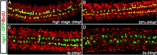
RB sensory neurons are born during gastrulation in zebrafish. A-D: Confocal micrographs of dorsal views of the spinal cord in 24-hpf Tg(neurogenin1:GFP) embryos, in which RB neurons express GFP. Embryos were treated with BrdU from high stage (A), 50% epiboly (B), tail bud (C), or 2 somites (D) through 24 hpf. In embryos treated from high stage or 50% epiboly (A,B), most of the RBs show BrdU incorporation, while in embryos in which BrdU exposure began at tail bud (C) or 2 somites (D), few RBs show BrdU incorporation. |
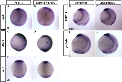
prdm1a affects dlx3b and dlx4b expression unidirectionally at the NPB. A,B: dlx3b expression is decreased at the NPB in prdm1a mutant embryos (B) compared to WT embryos (A) at tail bud. C-F: Embryos injected with antisense oligonucleotide morpholino to prdm1a show a decrease in dlx4b at the NPB at tail bud (D) and an absence of HuC-positive RBs (F) at 2 somites compared to un-injected controls (C, E). Conversely, prdm1a expression at the NPB shows no change in dlx3b/4b morpholino-injected embryos [compare un-injected controls at 90% epiboly (G) and tail bud (I) to morphants (H,J)]. Lateral (A-D, G-J) or dorsal (E,F) views with anterior toward the top. |
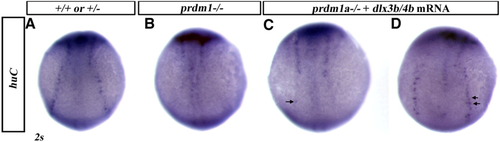
dlx3b and dlx4b function downstream of prdm1a to specify RB sensory neurons. Dorsal views with anterior toward the top. Wild type embryo (A) and prdm1a homozygous mutant embryos (B-D) stained for HuC, 2-3 somites. A,B: Un-injected dorsal views with anterior toward the top. Un-injected prdm1a mutants lack RBs at the NPB. C,D: mRNA for dlx3b and dlx4b partially rescues the RB phenotype to a varying degree in prdm1a mutants (arrows). |
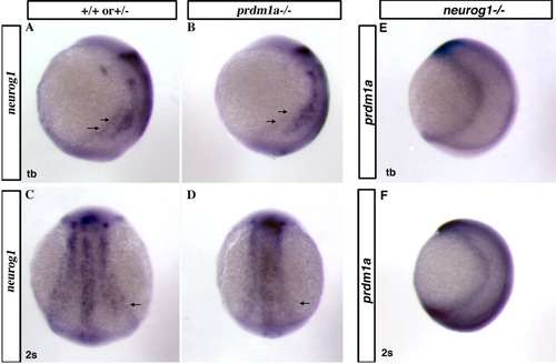
neurogenin1 expression is decreased at the NPB in prdm1a mutants. Lateral (A,B, E, F) or dorsal (C, D) views with anterior toward the top. neurogenin1 is expressed in three pairs of stripes at tail bud (A,B) and 2 somites (C,D). The most lateral stripes on either side of dorsal midline correspond to the NPB (arrows in A, C). This stripe of expression is reduced or missing in homozygous mutant prdm1a embryos (arrows in B, D). Conversely, prdm1a expression at tailbud and 2-somites stage is normal in neurogenin1(neurog1) mutant embryos (E,F). |
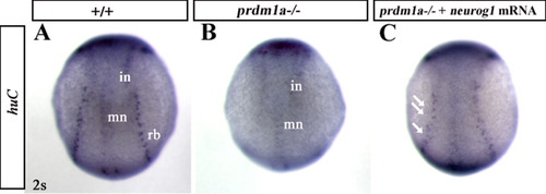
neurogenin1 functions downstream of prdm1a to specify RB sensory neurons. Dorsal views with anterior toward the top. Wild type embryo (A) and prdm1a homozygous mutant embryos (B, C) stained for HuC, 2-3 somites (A) HuC stains RBs, motoneurons (mn), and interneurons (in). B: Un-injected prdm1a mutants lack RBs at the NPB. C: mRNA for neurogenin1 partially rescues the RB phenotype to a varying degree in prdm1a mutants (arrows). |
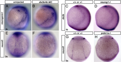
dlx3b and neurogenin1 do not regulate one another's transcription at the NPB. A-D: lateral views, anterior to the top. E-H: dorsal views, anterior to the top. Uninjected (A) and dlx3b/4b MO-injected (B) embryo at tailbud shows no difference in neurogenin1 expression at the NPB. Similarly, dlx3b expression is unchanged in neurogenin1 mutants (D) as compared to wildtype in (C) in tailbud stage embryos. neuroD is typically expressed at the NPB (E,G) but is absent or reduced in dlx3b/4b morphants (F) and prdm1a mutants (H). |