
Image
|
Figure Caption
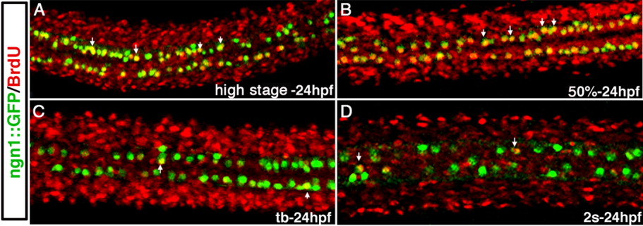
Fig. 3 RB sensory neurons are born during gastrulation in zebrafish. A-D: Confocal micrographs of dorsal views of the spinal cord in 24-hpf Tg(neurogenin1:GFP) embryos, in which RB neurons express GFP. Embryos were treated with BrdU from high stage (A), 50% epiboly (B), tail bud (C), or 2 somites (D) through 24 hpf. In embryos treated from high stage or 50% epiboly (A,B), most of the RBs show BrdU incorporation, while in embryos in which BrdU exposure began at tail bud (C) or 2 somites (D), few RBs show BrdU incorporation.
Acknowledgments
This image is the copyrighted work of the attributed author or publisher, and
ZFIN has permission only to display this image to its users.
Additional permissions should be obtained from the applicable author or publisher of the image.
Full text @ Dev. Dyn.