
|
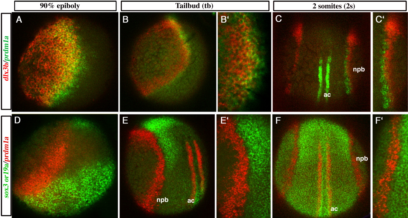
Fig. 2 Overlap of the prdm1a expression domain with dlx3b is greater than with neural markers sox3 and sox19a. Confocal micrographs of wild type embryos mounted to best observe potential overlap of expression at the NPB; (A-B′,D) mounted laterally with dorsal toward the right, (C-C′,F-F′) mounted dorsally with anterior toward the top, and (E-E′) mounted dorsolaterally with anterior toward the top. A-C′: prdm1a in green, dlx3b in red, overlap in yellow. D: prdm1a in green, sox3 in red, overlap in yellow. E-F′: prdm1a in red, sox19a in green. The prdm1a domain overlaps extensively with dlx3b at 90% epiboly (yellow in A), with less overlap at tail bud (B,B′), and very little overlap by 2 somites (C,C′). D: A small amount of overlap of prdm1a and sox3 is observed at 90% epiboly (yellow). Transcripts for sox19a (green) and prdm1a (red) abut one another, but do not overlap with each other, at tail bud (E-E′) and 2 somites (F-F′). This is especially evident at 20x (E′,F′). prdm1a is also expressed in the adaxial cells in the mesoderm near dorsal midline at tail bud and 2 somites. ac, adaxial cells, npb, neural plate border. A-F, 10x; B′-F′, 20x.