
- Title
-
Afferent neurons of the zebrafish lateral line are strict selectors of hair-cell orientation
- Authors
- Faucherre, A., Pujol-Martí, J., Kawakami, K., and Lopez-Schier, H.
- Source
- Full text @ PLoS One
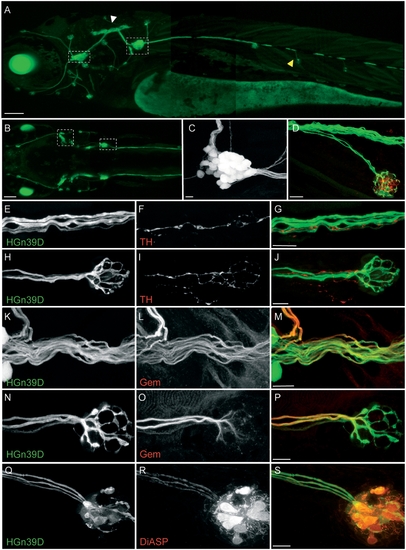
HGn39D transgenic zebrafish express GFP in the lateral line afferent neurons. (A–C) GFP expression pattern in a 4 dpf HGn39D transgenic fish. Maximal projections of lateral- (A) and top-views (B) are depicted. White and yellow arrowheads point to the neuronal projections to the hindbrain and to the neuromast respectively. Dashed frames indicate the lateral line ganglia in A and B. (C) Maximal projections of the posterior lateral line ganglion; approximately 25 somata can be counted. (D) Maximal projection of an HGn39D innervated neuromast immunolabeled with anti-HCS1 antibody (red). (E–P) Anti- tyrosine hydroxylase (TH) and Gem25.2/Cav1.3a (Gem) immunostainings were performed on 7 dpf HGn39D fish. (E,H,K,N) HGn39D GFP expression; (F,I) anti-TH antibody labels the efferent neurons; (L,O) Anti-Gem antibody labels the afferent neurons. Maximal projections of the posterior lateral line nerve (E–G, K–M) and of the terminal neuromast of the posterior lateral line (H–J, N–P) are depicted. (G,J,M,P). Overlays of HGn39D GFP expression and specific immunostainings. (Q–S) DiASP is trans-synaptically transported into the HGn39D afferent neurons. (Q) HGn39D GFP expression; (R) DiASP incorporation; (S) Overlay of HGn39D GFP expression and DiASP incorporation. Scale bars: 100 μm (A, B) and 10 μm (C–S). |
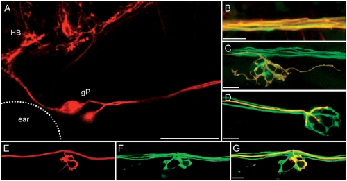
The HuC promoter drives the expression of mem-TdTomato in the afferent neurons. Maximal projections of HGn39D fish injected with HuC:mem-TdTomato are depicted. (A) HuC promoter drives the expression of mem-TdTomato in the soma of the afferent neurons (HB: hinbrain, gP: Posterior lateral-line ganglion). (B–C) mem-TdTomato is expressed in GFP-positive neurons of HGn39D fish at the level of the defasciculated fibers (B) and of the dendrites beneath the neuromast (C). (D) mem-TdTomato is expressed in a single neurons. (E–G) Injection of HuC:mem-TdTomato allows the mosaic expression of red neurons. A single red neuron (E) can be observed in HGn39D (F). (G) Overlay of HGn39D GFP and mem-TdTomato. Scale bars: 50 μm (A) and 10 μm (B–G). |
Afferent neurons innervate hair cells of identical polarity. Top views of neuromasts from HuC:mem-TdTomato injected ET4 fish are depicted. (A–D) maximal projections (A,C) and phalloidin staining (B,D) of neuromasts innervated by an afferent neuron that exclusively innervates hair cells of the same polarity (A–B) and by an afferent neuron that also projects neurites to hair cells of the opposite polarity (C–D). Asterisks indicate the innervated hair cells. Double asterisks point the innervated hair cells of the opposite polarities than the hair cells marked with single asterisks. (E–L) Maximal projection (E–K) and phalloidin staining (L) of the same neuromast. White arrows indicate a non-stable neurite contacting hair cells of opposite polarity (double asterisks) compared to stable synapses (asterisks). Scale bars: 10 μm (A,C, E and K) and 5 μm (B, D and L). |
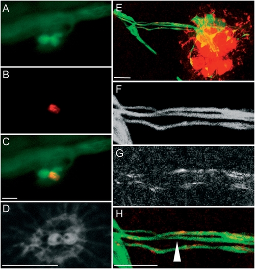
Polarized fluid jet induces incorporation of DiASP in hair cells of selected orientation and is transported to a subset of the afferent neurons. (A–D) DiASP directional incorporation in a neuromast with two hair cells of opposite polarities. (A) GFP expression in HGn39D/SqET4 fish; (B) DiASP incorporation; (C) overlay of GFP expression and DiASP; (D) phalloidin staining reveals the orientation of the hair cells. (E–H) DiASP directional incorporation in a fully developed neuromast doesn't label all the afferent neurons. (E) overlay of HGn39D GFP expression and DiASP incorporation in the hair cells and the afferent neurons ; (F–H) details of the neurons with GFP expression (F), DiASP (G) and overlay of the two (H). The white arrow indicates an afferent neuron that is not labeled by the DiASP. Scale bar: 10 μm. |
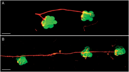
Afferent innervation of adjacent neuromasts. (A–B) Maximal projections of single neurons labelled with mem-TdTomato innervating hair cells of the same polarity of (A) two adjacent neuromasts in an SqET4 transgenic fish and (B) three adjacent neuromasts in a brn3C:GFP transgenic fish. Those depicted are the terminal neuromasts from a 4 dpf fish. Scale bar: 20 μm. EXPRESSION / LABELING:
|
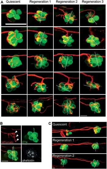
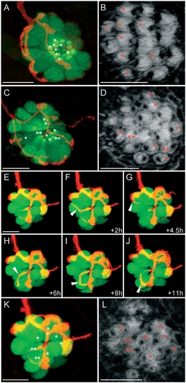
Afferent neurons reinnervate the hair cells of the same polarity after several cycles of ablation and regeneration. (A) Maximal projections of SqET4 (two first lines) or brn3c:GFP fish neuromasts innervated by a single neuron expressing mem-TdTomato. Confocal z-stacks of the same neuromast have been acquired at 3 dpf (quiescent state, first row) and 20 hours after each of three rounds of neomycin treatments (Regeneration 1, 2 and 3). All rows picture neuromasts with parallel cellular polarity (hair bundles aligned with the anteroposterior axis of the fish body) except the penultimate, which shows a neuromast with perpendicular cellular polarity. (B) Phalloidin staining performed on the last neuromast depicted on A after the third regeneration process. (C) Maximal projections of two brn3c:GFP adjacent neuromasts innervated by a single neuron expressing mem-TdTomato. Confocal z-stacks of the same neuromasts have been acquired at 3 dpf (quiescent) and 20 hours after each of two rounds of neomycin treatments (Regeneration 1 and 2). Dotted lines represent the axis of polarity of the neuromast. White arrowheads in panel B indicate the bulged neurites establishing contacts with the two hair cells of same polarity. Scale bar: 10 μm. |
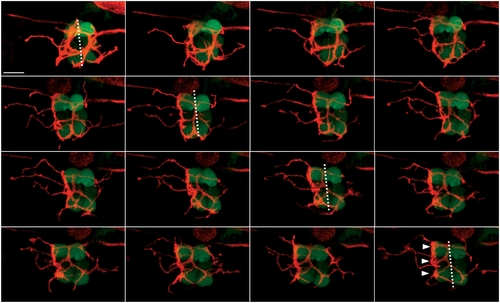
Afferent innervation of regenerating hair cells is a dynamic process. Maximal projections of an SqET4 fish neuromast innervated by an afferent neuron expressing mem-TdTomato. Pictures have been taken every 45 min, starting 20 hours after neomycin treatment. Dotted lines represent the axis of polarity of the neuromast. White arrowheads point to the bulged neurites establishing contacts with the three hair cells of same polarity. Scale bar: 10 μm. |