
Fig. 6
- ID
- ZDB-FIG-090309-11
- Publication
- Faucherre et al., 2009 - Afferent neurons of the zebrafish lateral line are strict selectors of hair-cell orientation
- Other Figures
- All Figure Page
- Back to All Figure Page
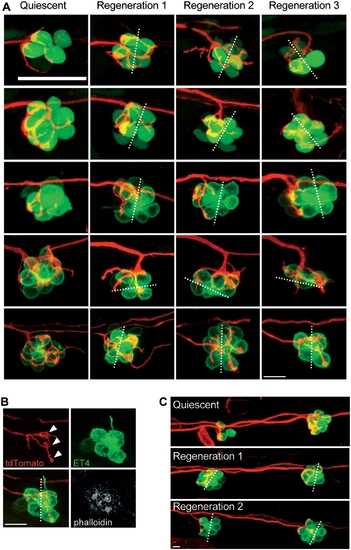
Afferent neurons reinnervate the hair cells of the same polarity after several cycles of ablation and regeneration. (A) Maximal projections of SqET4 (two first lines) or brn3c:GFP fish neuromasts innervated by a single neuron expressing mem-TdTomato. Confocal z-stacks of the same neuromast have been acquired at 3 dpf (quiescent state, first row) and 20 hours after each of three rounds of neomycin treatments (Regeneration 1, 2 and 3). All rows picture neuromasts with parallel cellular polarity (hair bundles aligned with the anteroposterior axis of the fish body) except the penultimate, which shows a neuromast with perpendicular cellular polarity. (B) Phalloidin staining performed on the last neuromast depicted on A after the third regeneration process. (C) Maximal projections of two brn3c:GFP adjacent neuromasts innervated by a single neuron expressing mem-TdTomato. Confocal z-stacks of the same neuromasts have been acquired at 3 dpf (quiescent) and 20 hours after each of two rounds of neomycin treatments (Regeneration 1 and 2). Dotted lines represent the axis of polarity of the neuromast. White arrowheads in panel B indicate the bulged neurites establishing contacts with the two hair cells of same polarity. Scale bar: 10 μm. |