
- Title
-
Neuropilin-1 Modulates p53/Caspases Axis to Promote Endothelial Cell Survival
- Authors
- Wang, L., Dutta, S.K., Kojima, T., Xu, X., Khosravi-Far, R., Ekker, S.C., and Mukhopadhyay, D.
- Source
- Full text @ PLoS One
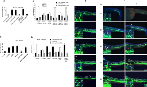
NRP-1 mediates VPF/VEGF-induced EC survival; the C-terminal three amino acids of NRP-1 are essential for this function. Quantitative determination of apoptotic nuclei in PAEC (n = 8). TUNEL assay was performed in apoptosis-induced PAEC and PAEC/NRP-1 with or with out 10ng/ml VPF/VEGF stimulation for 48 hrs to assess whether NRP-1 mediates PAEC survival. b. FACS apoptosis analysis to determine the percentage of apoptotic PAEC. c. Quantitative determination of apoptotic nuclei in HUVEC (n = 8. n: The number of counted field. All are same in this study). EGNP-1 mediates HUVEC survival; the C-terminal three amino acids of NRP-1 are essential for this function. TUNEL assay was performed in apoptosis-induced HUVEC transfected with EGNP-1 or EGNP-1&detla;SEA and stimulated with or without 10ng/ml EGF. d. FACS apoptosis analysis to determine the percentage of apoptotic HUVEC. All the experiments at least were repeated three times and duplicated-wells were used for each experiment. *p<0.001, **p<0.005, ***p<0.01, ****p<0.05 in a Student's t test. e. NRP-1 mediates EC survival in zebrafish; the C-terminal three amino acids (SEA-COOH) are essential for the function. Fluorescent image of embryos injected with indicated morpholinos and mRNA, then subjected to the antibody stain with anti-GFP antibody and TUNEL assay to detect apoptosis. Insets show a magnification of apoptotic foci in the vessel. The arrows indicate apoptotic EC. i. Control; ii. Mismatch MO (4.5 ng); iii. zNRP-1a/1b MOs (4.5 ng+4.5 ng); iv. zNRP-1a/1b MOs (4.5 ng+4.5 ng)+hNRP-1 mRNA (0.3ng); v. zNRP-1a/1b MOs (4.5ng+4.5ng)+hNRP-1&detla;SEA mRNA (0.3 ng). f. The dynamics of NRP-1-induced EC apoptosis in the zebrafish embryo. Fluorescent images of different stage embryos injected with indicated morpholinos and subjected to the antibody stain and TUNEL assay to detect apoptosis. Insets show a magnification of apoptotic foci in the vessel. The arrows indicate apoptotic EC. i. Control; ii. zNRP-1a/1b MOs (3.0 ng+3.0 ng). |
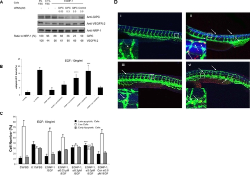
GIPC is involved in NRP-1-mediated EC survival. Knockdown of GIPC by siRNA in HUVEC. HUVEC were transfected with siRNA against GIPC in different concentrations. Immunoblotting was performed to quantify the expression levels of GIPC in these cells. GIPC and VEGFR-2 were quantified as a ratio to the amount of NRP-1. b. GIPC is involved in NRP-1-mediated HUVEC survival. Apoptosis assays were performed in apoptosis-induced HUVEC transfected with EGNP-1 and then transfected with siRNA, and followed by 10 ng/ml EGF stimulation for 48 hours. Quantitative determination of apoptotic nuclei (n = 8). Knock down of GIPC inhibits EGF-stimulated HUVEC apoptosis in a dosage-dependent manner. c. FACS apoptosis analysis to assess the percentage of apoptotic HUVEC. All the experiments at least were repeated three times and duplicated-wells were used for each experiment. *p<0.001, **p<0.005, ***p<0.01, ****p<0.05 in a Student's t test. d. GIPC is involved in NRP-1-mediated EC survival in zebrafish. Fluorescent image of embryos injected with indicated morpholinos and mRNA, then subjected to the antibody stain with anti-GFP antibody and TUNEL assay to detect apoptosis. Insets show a magnification of apoptotic foci in the vessel. The arrows indicate apoptotic EC. i. Control; ii. zGIPC MO (4.5 ng); iii. zGIPC (4.5 ng)+hGIPC mRNA (0.3 ng); iv. zGIPC MO (4.5 ng)+hGIPCδPDZ mRNA (0.3 ng). |
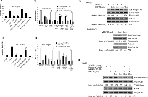
PI-3K/Akt is involved in NRP-1-mediated HUVEC survival. PI3K is involved in NRP-1-mediated EC survival signaling. Apoptosis assay was performed in apoptosis-induced HUVEC transfected with EGNP-1 or co-transfected with EGNP-1 and p85(DN) and stimulated with or without 10ng/ml EGF for 48hours, and then treated with LY294002 for 30 minutes, accordingly. Apoptotic nuclei were determined quantitatively (n = 8). b. FACS apoptosis analysis to quantify the percentage of apoptotic HUVEC. c. Akt is involved in PI3K-mediated NRP-1 survival signaling in EC. Apoptosis assays were performed in apoptosis-induced HUVEC transfected with EGNP-1 or co-transfected with EGNP-1 and Akt(DN) or Akt(Active) and stimulated with or without 10ng/ml EGF. Apoptotic nuclei were determined quantitatively (n = 8). d. FACS apoptosis analysis to determine the percentage of apoptotic HUVEC. All the experiments at least were repeated three times and duplicated-wells were used for each experiment. *p<0.001, **p<0.005, ***p<0.01 in a Student's t test. e. Akt phosphorylation by NRP-1/EGNP-1 activation. HUVEC were transfected with EGNP-1 and then stimulated with EGF. Immunoblotting with anti-phospho-Akt was carried out to quantify the expression levels of phospho-Akt in these cells. β-Actin was used as a loading control f. The influence of PI3K depletion and VEGFR-2 phosphorylation inhibition on EGNP-1-mediated Akt activation. Immunoblotting was performed in HUVEC transfected with EGNP-1 and then treated with Ly294002 or VEGFR-2 Kinase inhibitor IV. Phospho-Akt and Akt were quantified as a ratio to the amount of β-Actin. |
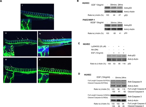
p53 inactivation in NRP-1-mediated EC survival. p53 is involved in NRP-1-mediated EC survival in zebrafish. Fluorescent image of embryos injected with indicated morpholinos and subjected to the antibody stain with anti-GFP antibody and TUNEL assay to detect apoptosis; Insets show a magnification of apoptotic foci in the vessel. The arrows indicate apoptotic EC. i. Control; ii. zNRP-1a/1b MOs (4.5 ng+4.5 ng); iii. zNRP-1a/1b MOs (4.5 ng+4.5 ng)+zp53 MO (9.0 ng); iv. zMdm2 MO (4.5ng); v. zMdm2 MO (4.5 ng)+zp53 MO (9.0 ng). b. NRP-1/EGNP-1-mediated p53 inactivation. EGNP-1 transduced HUVEC or PAEC/NRP-1 were subjected to EGF or VEGF for the indicated times and their lysates were immunoblotted for p53 with β-actin as a loading control. c. p53 is the downstream signaling molecule of PI3K/Akt in EGNP-1-mediated EC survival. HUVEC were transfected with EGNP-1 then treated with Ly294002 or co-transfected with EGNP-1 and Akt(DN). Immunoblotting was performed to determine p53 expression level. d. Inhibition of mitochondrial apoptotic pathway in EGNP-1-mediated EC survival. Immunoblotting was carried out in EGNP-1 transduced HUVEC stimulated with EGF at indicated times to assess the activation of caspase-9 and caspase-3. p53 and caspases were quantified as a ratio to the amount of β-Actin *p<0.001, **p<0.005, ***p<0.01, ****p<0.05 in a Student's t test. |
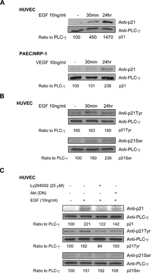
Induction of p21 through PI3K/Akt is involved in the survival signaling of NRP-1. The activation of NRP-1/EGNP-1 increased p21 expression. EGNP-1 transduced HUVEC or PAEC/NRP-1 were subjected to EGF or VEGF for the indicated times and their lysates were immunoblotted for p21 with PLC-γ as a loading control. b. The activation of EGNP-1 induced p21 phosphorylation at its Tyr145 and Ser146 sites. Immunoblotting was performed with anti-p21Tyr and anti-p21Ser in EGNP-1 transduced HUVEC stimulated by EGF for the indicated times. c. Akt mediates the phosphorylation of p21 in EGNP-1-mediated signaling. HUVEC were transfected with EGNP-1 then treated with Ly294002 or co-transfected with EGNP-1 and Akt(DN). Immunoblotting was performed to quantify the expression and activation of p21. p21 and phospho-p21 were quantified as a ratio to the amount of PLC-γ. *p<0.001, **p<0.005, ***p<0.01, ****p<0.05 in a Student's t test. |
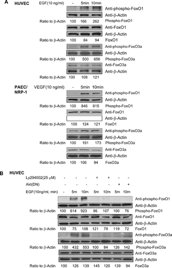
Involvement of transcription factors FoxO1 and FoxO3a in NRP-1-mediated EC survival signaling. Phosphorylation of FoxO1 and FoxO3a in NRP-1 survival signaling. After EGF stimulation of EGNP-1-transduced HUVEC or VEGF stimulation of PAEC/NRP-1 for the indicated times, the protein levels in cell extracts of FoxO1 and FoxO3a were assessed by immunoblotting. b. PI3K/Akt depletion resulted in reduction of FoxO1 and FoxO3a phosphorylation. HUVEC were transfected with EGNP-1, then treated with Ly294002 or co-transfected with EGNP-1 and Akt(DN). Immunoblotting was performed to determine the phosphorylation level of FoxO1 and FoxO3a with β-Actin as a loading control. phospho-FoxOs and FoxOs were quantified as a ratio to the amount of β-Actin. *p<0.001, **p<0.005, ***p<0.01, ****p<0.05 in a Student's t test. |