
|
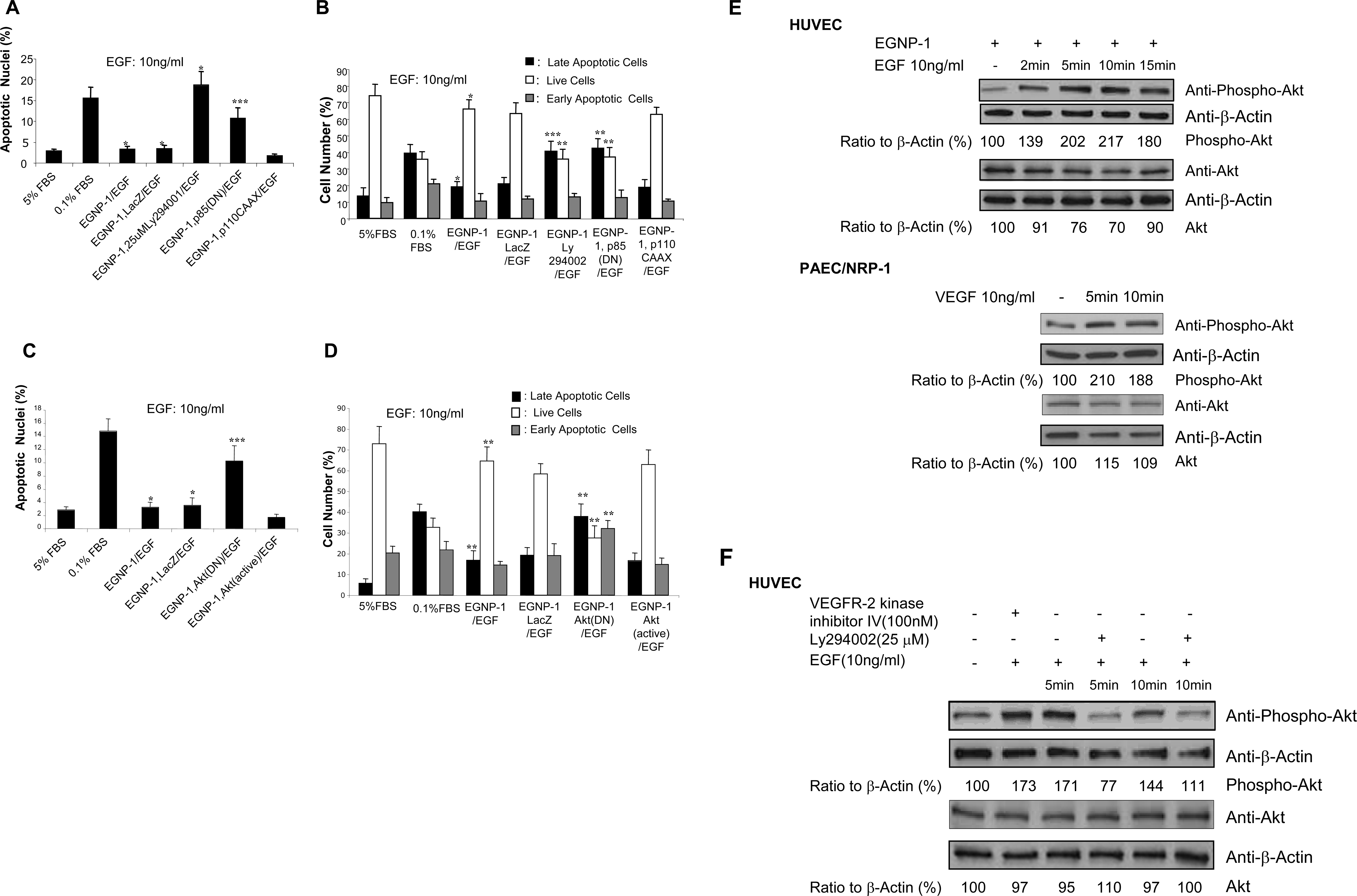
Fig. 3 PI-3K/Akt is involved in NRP-1-mediated HUVEC survival. PI3K is involved in NRP-1-mediated EC survival signaling. Apoptosis assay was performed in apoptosis-induced HUVEC transfected with EGNP-1 or co-transfected with EGNP-1 and p85(DN) and stimulated with or without 10ng/ml EGF for 48hours, and then treated with LY294002 for 30 minutes, accordingly. Apoptotic nuclei were determined quantitatively (n = 8). b. FACS apoptosis analysis to quantify the percentage of apoptotic HUVEC. c. Akt is involved in PI3K-mediated NRP-1 survival signaling in EC. Apoptosis assays were performed in apoptosis-induced HUVEC transfected with EGNP-1 or co-transfected with EGNP-1 and Akt(DN) or Akt(Active) and stimulated with or without 10ng/ml EGF. Apoptotic nuclei were determined quantitatively (n = 8). d. FACS apoptosis analysis to determine the percentage of apoptotic HUVEC. All the experiments at least were repeated three times and duplicated-wells were used for each experiment. *p<0.001, **p<0.005, ***p<0.01 in a Student's t test. e. Akt phosphorylation by NRP-1/EGNP-1 activation. HUVEC were transfected with EGNP-1 and then stimulated with EGF. Immunoblotting with anti-phospho-Akt was carried out to quantify the expression levels of phospho-Akt in these cells. β-Actin was used as a loading control f. The influence of PI3K depletion and VEGFR-2 phosphorylation inhibition on EGNP-1-mediated Akt activation. Immunoblotting was performed in HUVEC transfected with EGNP-1 and then treated with Ly294002 or VEGFR-2 Kinase inhibitor IV. Phospho-Akt and Akt were quantified as a ratio to the amount of β-Actin.