
|
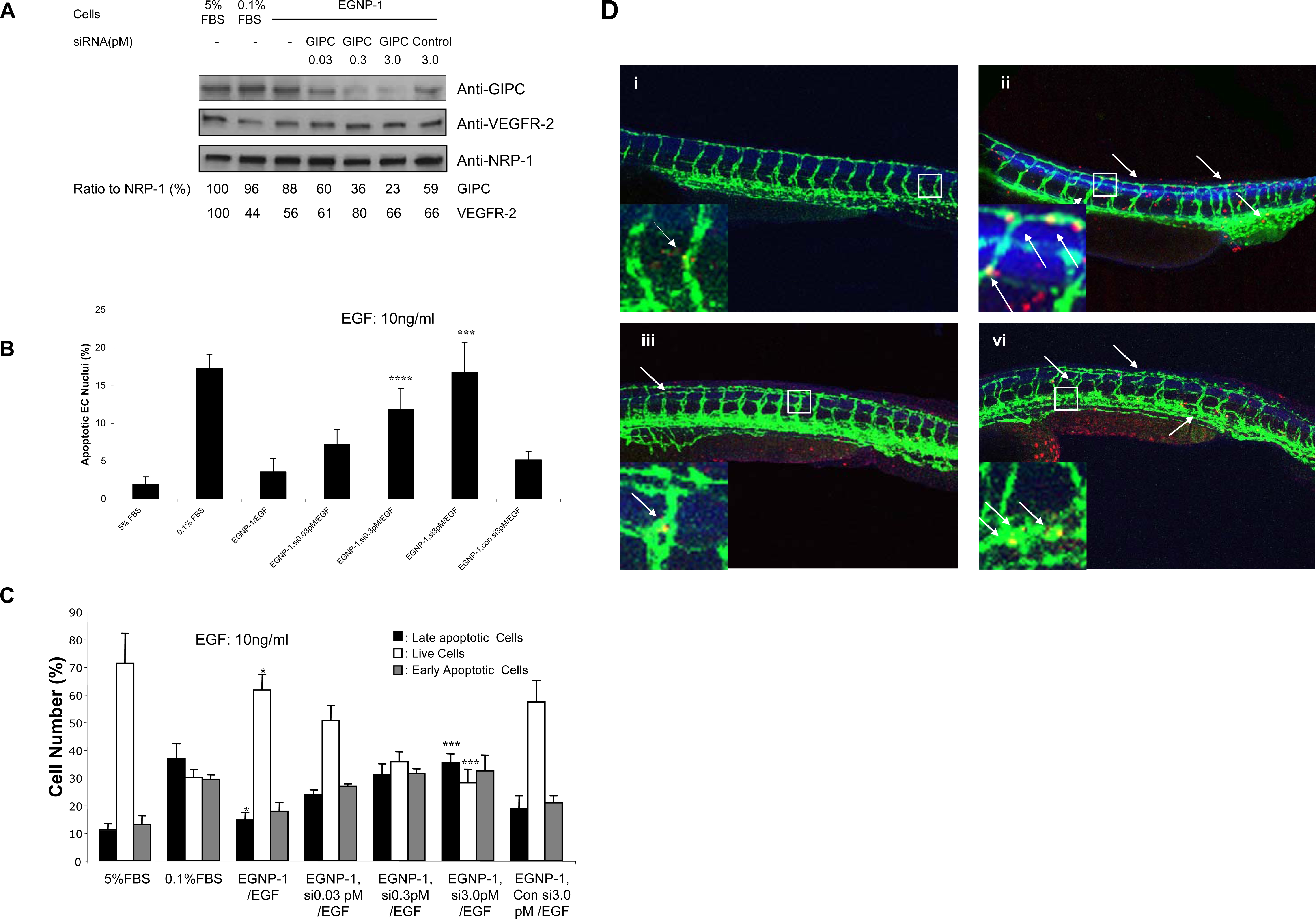
Fig. 2 GIPC is involved in NRP-1-mediated EC survival. Knockdown of GIPC by siRNA in HUVEC. HUVEC were transfected with siRNA against GIPC in different concentrations. Immunoblotting was performed to quantify the expression levels of GIPC in these cells. GIPC and VEGFR-2 were quantified as a ratio to the amount of NRP-1. b. GIPC is involved in NRP-1-mediated HUVEC survival. Apoptosis assays were performed in apoptosis-induced HUVEC transfected with EGNP-1 and then transfected with siRNA, and followed by 10 ng/ml EGF stimulation for 48 hours. Quantitative determination of apoptotic nuclei (n = 8). Knock down of GIPC inhibits EGF-stimulated HUVEC apoptosis in a dosage-dependent manner. c. FACS apoptosis analysis to assess the percentage of apoptotic HUVEC. All the experiments at least were repeated three times and duplicated-wells were used for each experiment. *p<0.001, **p<0.005, ***p<0.01, ****p<0.05 in a Student's t test. d. GIPC is involved in NRP-1-mediated EC survival in zebrafish. Fluorescent image of embryos injected with indicated morpholinos and mRNA, then subjected to the antibody stain with anti-GFP antibody and TUNEL assay to detect apoptosis. Insets show a magnification of apoptotic foci in the vessel. The arrows indicate apoptotic EC. i. Control; ii. zGIPC MO (4.5 ng); iii. zGIPC (4.5 ng)+hGIPC mRNA (0.3 ng); iv. zGIPC MO (4.5 ng)+hGIPCδPDZ mRNA (0.3 ng).