FIGURE SUMMARY
Title

MicroRNAs show a wide diversity of expression profiles in the developing and mature central nervous system

Authors
Kapsimali, M., Kloosterman, W.P., de Bruijn, E., Rosa, F., Plasterk, R.H., and Wilson, S.W.
Source
Full text @ Genome Biol.

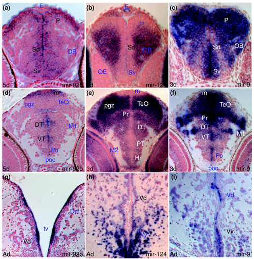
miRNAs expressed in proliferating and/or differentiating cells in the developing and adult zebrafish brain. In this and other figures, unless otherwise mentioned, sections are transverse with dorsal on the top, stage is shown bottom left and miRNA analyzed by in situ hybridization bottom right, in situ staining is in blue and cell nuclei are visualized with nuclear red counterstaining. Abbreviations used in the Results section of the text are denoted in black. For other abbreviations, see Additional data file 26. (a,d,g) miR-92b expression in periventricular and adjacent cells of the telencephalon (a,g), diencephalon and optic tectum (d). (b,e,h) miR-124 expression in differentiating cells in the telencephalon (b,h), diencephalon and optic tectum (e). (c,f,i) miR-9 expression in periventricular/proliferating and differentiating cells of the telencephalon (c,i), diencephalon and optic tectum (f).

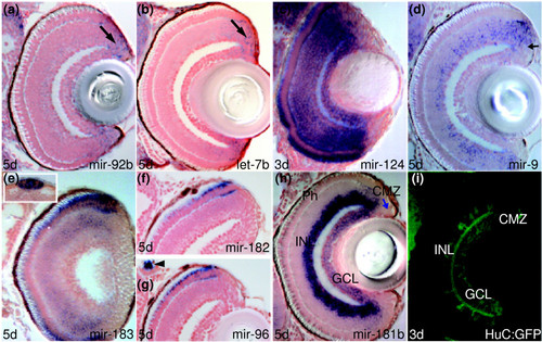
Several miRNAs expressed in discrete retinal cell populations. (a-h) Transverse sections through retinae in situ hybridized with miR-92b, let-7b, miR-124, miR-9, miR-183, miR-182, miR-96 and miR-181b probes. Arrows point at proliferative ciliary marginal zone (CMZ) cells in (a,b,d,h). The inset in (e) shows pineal cells. The arrowhead in (g) indicates miR-96 expression in peripheral sensory neuromasts. (i) Confocal section through the retina of a transgenic line Tg(huC:GFP) immunostained for GFP. Other miRNAs with expression in the retina include miR-454a (Figure C in Additional data file 25), miR-132 (Figure E in Additional data file 25), miR-125b (Figure F in Additional data file 25) and miR-181a (Figure G in Additional data file 13).

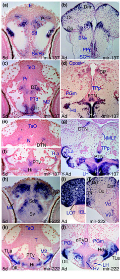
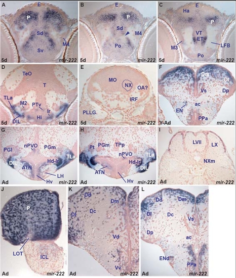
miR-137 and miR-222 expression is conserved between larval and adult brain. (a,c,e,f) miR-137 expression in the larval caudal telencephalon (a), diencephalon (c), dorsal midbrain (e) and hypothalamus (f). (b,d,g) miR-137 expression in adult brain sections at levels corresponding to the embryonic sections shown in (a), (c) and (e/f), respectively. (h,k) miR-222 expression in the larval telencephalic pallium (P) and subpallium (Sd, Sv), hypothalamus (Hi, TLa, DIL, lr) and posterior tuberculum (PTv, M2). (i,j,l) miR-222 expression in corresponding adult nuclei in the pallium (P, Dm, Dl, Dd, Dc), subpallium (Vd, Vv), hypothalamus (ATN, LH, TLa, DIL, Hd-lr) and posterior tuberculum (nPVO, PGl).

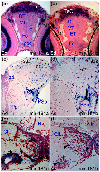
Conserved and divergent expression of miR-181a and miR-181b. (a,b) miR-181a and miR-181b expression in larval tectal (TeO) and migrated pretectal area cells (M1). (c,d) Comparable miR-181a and miR-181b expression in the adult optic tectum (sgz, cz) and pretectal nuclei (PSm, PSp). (e) miR-181a is expressed in more cells than (f) miR-181b (arrowheads) in the adult hypothalamic mamillary body (CM) and dorsal periventricular zone (Hd).

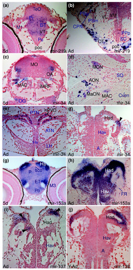
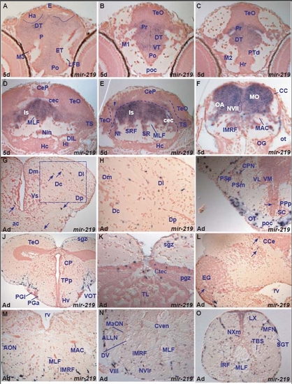
Examples of miRNAs showing differences in expression between larval and adult stages. (a,b) miR-219 expression in the diencephalon at the level of the post-optic commissure (poc) of the larval and adult brain. In the larval brain (a), miR-219 is widely expressed in the ventral (VT) and dorsal (DT) thalamus and periventricular pretectum (Pr) whereas cells of the poc are devoid of expression. In contrast, in the adult (b), miR-219 is expressed in cells in the poc and optic tract (OT) whereas ventrolateral (VL) and ventromedial (VM) thalamic nuclei are devoid of expression. (c-f) miR-34 expression: (c,d) show conserved miR-34 expression in the octaval area (OA, MON, AON, MaON) and Mauthner neuron (MAC) in the larval and adult zebrafish brain, respectively; (e) shows miR-34 in the adult nucleus of the paraventricular organ (nPVO) and the arrow in (f) points to miR-34 expressing cells in the lateral part of the adult left habenula. (g,h) Conserved miR-153a expression throughout the larval (Ha) and adult (Hav, Had) habenulae. (i) miR-137 expression in groups of dorsal habenular cells (Had, arrowheads). (j) miR-138 expression in groups of dorsal habenular cells (Had).

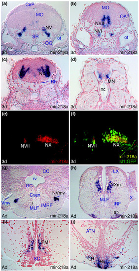
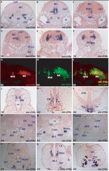
miR-218a is expressed in embryonic cranial and spinal motor-neurons. (a-d,g-h) Larval and adult miR-218a expression in the motor nuclei of the fifth (NV, NVmv), sixth (NVI), seventh (NVII), tenth (NX, NXm) cranial nerves and spinal motor neurons (MN). (e,f) Confocal sagittal sections through the hindbrain of an embryo expressing the Tg(isl1:GFP) transgene with anterior to the left. miR-218a expression is shown in red in (e) and (f) is a superimposition of the miR-218a staining (red) and anti-GFP immunostaining (green). Yellow cells express both miR-218a and GFP in the NVII and NX cranial motor nuclei. (h-j) Additional sites of expression of miR-218a in the adult inferior olive (IO), preoptic magnocellular area (PM) and hypothalamus (Hv, LH).

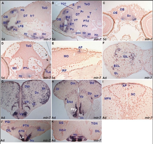
miR-7 expression in the zebrafish brain. miR-7 shows spatially localized expression, largely conserved throughout life. Larval expression is restricted to the forebrain in telencephalic, preoptic, few thalamic, eminentia thalami and ventral hypothalamic cells. In addition, it is expressed in a few cells of the area postrema at the dorsal junction of hindbrain-spinal cord (Table D). miR-7 expression is mainly conserved in telencephalic, hypothalamic nuclei and area postrema between the larval and adult zebrafish brain. However, in the adult, we observe expression in the external and internal cellular layers of the olfactory bulb. Furthermore, in contrast to the larval, the adult thalamus is devoid of miR-7 expression (Table D, I). miR-7 often shares expression in the forebrain with miR-222 but there are also some differences (table D, I).

A. parasagittal section through the larval fore- and midbrain showing miR-7 expressing cells in the hypothalamus (caudal, Hc, intermediate, Hi and rostral, Hr), preoptic area (Po), eminentia thalami (ET), ventral subpallium (Sv) around the anterior commissure and pallium (P).
B. sagittal section through the larval fore- and midbrain showing miR-7 expressing cells in the hypothalamus (caudal, Hc, intermediate, Hi and rostral, Hr), preoptic area (Po), eminentia thalami (ET), ventral (Sv) and dorsal (Sd) subpallium and pallium (P).
C. transverse section through the rostral larval telencephalon showing miR-7 expressing cells in the ventral subpallium (Sv).
D. transverse section through the larval hypothalamus and midbrain showing miR-7 expressing cells in the rostral ventral hypothalamus (Hr).
E. sagittal section through the larval hindbrain showing miR-7 expressing cells in the caudal medulla oblongata (MO) at the level of the area postrema (AP).
F. transverse section through the adult olfactory bulb showing miR-7 expressing cells in the internal (ICL) and external (ECL) cellular olfactory layers.
G. transverse section through the adult telencephalon showing miR-7 expressing cells in the ventral (Vv), central (Vc) and dorsal (Vd) nuclei of the ventral telencephalon (subpallium), medial (Dm), central (Dc) and lateral (Dl) zones of the dorsal telencephalon (pallium). Expression is largely absent from cells lining the ventricles.
H. transverse section through the caudal adult telencephalon and rostral diencephalon (at the level of the optic chiasma, oc) showing miR-7 expressing cells in the suprachiasmatic nucleus (SC), posterior parvocellular preoptic nucleus (PPp), dorsal endopeduncular nucleus (ENd, part of the eminentia thalami), posterior nucleus of the ventral telencephalon (Vp), posterior (Dp), medial (Dm), dorsal (Dd) and lateral (Dl) zones of the dorsal telencephalon.
I. transverse section through the caudal medulla oblongata (MO) showing miR-7 expressing cells in the area postrema (AP).
J. transverse section through the adult hypothalamus showing miR-7 expressing cells in the lateral hypothalamic nucleus (LH), ventral zone of the periventricular hypothalamus (Hv), dorsal zone of the periventricular hypothalamus at the level of the lateral hypothalamic ventricular recess (Hd-lr) and diffuse nucleus of the inferior hypothalamic lobe (DIL).
K. transverse section through the adult hypothalamus (caudal to section J) showing miR-7 expressing cells in the caudal zone of the periventricular hypothalamus (Hc).

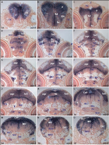
miR-9 expression in the 3dpf zebrafish brain.
miR-9 is expressed in both periventricular and adjacent cells of proliferative zones and differentiating cells arising from these domains. miR-9 expressing cells are widespread in all brain subdivisions (fore-, mid- and hind-brain) although some areas such as the larval epithalamus and the hypothalamic lateral torus are devoid of expression (Table A). In addition to the brain, miR-9 is expressed in the inner retinal nuclear layer and ciliary marginal zone. miR-9 expression is highly conserved between larval and adult brain and we find only minor differences at the regional level (Table A,F). miR-9 is expressed in a few adult habenular and hypothalamic lateral torus cells, areas devoid of larval expression.

A. transverse section through the larval rostral telencephalon at the level of the olfactory epithelium (OE) showing miR-9 expressing cells in the olfactory bulb (OB). Expression is absent in cells immediately ventral to the bulb.
B. transverse section through the larval telencephalon showing miR-9 expressing cells in the ventral (Sv) and dorsal (Sd) subpallium, pallium (P) and olfactory bulb (OB).
C. transverse section through the larval caudal telencephalon and epithalamus showing miR-9 expressing cells in the ventral (Sv) and dorsal (Sd) subpallium, pallium (P) and migrated telencephalic area (M4). Expression is absent from the habenulae and epiphysis.
D. transverse section through the larval diencephalon and rostral optic tectum showing miR-9 expressing cells in the preoptic area (Po), eminentia thalami (ET), migrated eminentia thalami (M3), ventral thalamus (VT), dorsal thalamus (DT), zona limitans intrathalamica (ZLI) and the optic tectum (TeO).
E. transverse section through the larval diencephalon and optic tectum showing miR-9 expressing cells in the preoptic area (Po), ventral thalamus (VT), dorsal thalamus (DT), periventricular (Pr) and migrated (M1) pretectal areas, optic tectum (TeO), retinal inner nuclear layer (INL) and ciliary marginal zone (arrowhead).
F. transverse section through the larval diencephalon and optic tectum showing miR-9 expressing cells in the rostral hypothalamus (Hr), periventricular posterior tuberculum (PT), ventral thalamus (VT), dorsal thalamus (DT), periventricular (Pr) and migrated (M1) pretectal areas, proliferative (m) and periventricular gray (pgz) zones of the optic tectum (TeO).
G. transverse section through the larval diencephalon and optic tectum showing miR-9 expressing cells in the rostral hypothalamus (Hr), ventral (PTv), dorsal (PTd) periventricular and migrated (M2) posterior tuberculum, dorsal thalamus (DT), periventricular pretectal area (Pr) and periventricular gray (pgz) zone of the optic tectum (TeO).
H. transverse section through the larval diencephalon and midbrain showing miR-9 expressing cells in the rostral hypothalamus (Hr), ventral (PTv), dorsal (PTd) periventricular and migrated (M2) posterior tuberculum, tegmentum (T), proliferative (m) and periventricular gray (pgz) zones of the optic tectum (TeO).
I. transverse section through the larval hypothalamus and midbrain showing miR-9 expressing cells in the intermediate hypothalamus (Hi, around the lateral ventricular recess, lr), ventral periventricular (PTv) and migrated (M2) posterior tuberculum, tegmental area at the level of the nucleus of the medial longitudinal fascicle (N), semicircular torus (TS) and periventricular gray zone (pgz) of the optic tectum (TeO).
J. transverse section through the larval hypothalamus and midbrain showing miR-9 expressing cells in the intermediate hypothalamus (Hi) at the level of the lateral hypothalamic ventricular recess (lr), tegmental area (T), occulomotor nucleus (NIII), semicircular torus (TS) and periventricular gray zone (pgz) of the optic tectum (TeO). The arrowhead points at the oculomotor nerve (III).
K. transverse section through the larval caudal hypothalamus and midbrain showing miR-9 expressing cells at the level of the lateral hypothalamic ventricular recess (lr), caudal hypothalamus (Hc), tegmental area (T), semicircular torus (TS), periventricular gray zone (pgz) of the optic tectum (TeO) and cerebellar valvula (Va).
L. transverse section through the larval caudal hypothalamus, midbrain and isthmus showing miR-9 expressing cells in the interpeduncular nucleus (NIn), isthmic area (Is), semicircular torus (TS), cerebellar plate (CeP) and optic tectum (TeO). M. transverse section through the larval hindbrain at the level of the octaval ganglion (OG) showing miR-9 expressing cells in the medulla oblongata (MO) and cerebellar plate (CeP).
N. transverse section through the larval hindbrain at the level of the otic capsule (caudal to section M) showing miR-9 expressing cells in the medulla oblongata (MO), presumptive octaval area (OA?) and rhombic lip (RL).
O. transverse section through the larval hindbrain at the level of the otic capsule (caudal to section N) showing miR-9 expressing cells in the medulla oblongata (MO), presumptive octaval area (OA?) and rhombic lip (RL).

miR-9 expression in the 5dpf zebrafish brain.
miR-9 is expressed in both periventricular and adjacent cells of proliferative zones and differentiating cells arising from these domains. miR-9 expressing cells are widespread in all brain subdivisions (fore-, mid- and hind-brain) although some areas such as the larval epithalamus and the hypothalamic lateral torus are devoid of expression (Table A). In addition to the brain, miR-9 is expressed in the inner retinal nuclear layer and ciliary marginal zone. miR-9 expression is highly conserved between larval and adult brain and we find only minor differences at the regional level (Table A,F). miR-9 is expressed in a few adult habenular and hypothalamic lateral torus cells, areas devoid of larval expression.

A. transverse section through the larval rostral telencephalon at the level of the olfactory epithelium (OE) showing miR-9 expressing cells in the olfactory bulb (OB).
B. transverse section through the larval telencephalon at the level of the anterior commissure (ac) showing weakly miR-9 expressing cells in the preoptic area (Po), dorsal subpallium (Sd), migrated telencephalic area (M4) and pallium (P).
C. transverse section through the larval caudal telencephalon and rostral diencephalon showing miR-9 expressing cells in the preoptic area (Po), eminentia thalami (ET), migrated eminentia thalami (M3) and pallium (P).
D. transverse section through the larval diencephalon and rostral optic tectum showing miR-9 expressing cells in the preoptic area, (Po), eminentia thalami (ET), migrated eminentia thalami (M3), ventral thalamus (VT), dorsal thalamus (DT), zona limitans intrathalamica (ZLI) and the optic tectum (TeO).
E. transverse section through the larval diencephalon and optic tectum showing miR-9 expressing cells in the intermediate hypothalamus (Hi), ventral (PTv), dorsal (PTd) periventricular and migrated (M2) posterior tuberculum, dorsal thalamus (DT), periventricular pretectal area (Pr), proliferative zone (m), periventricular gray (pgz) zone and longitudinal torus (TL) of the optic tectum (TeO).
F. transverse section through the larval caudal hypothalamus and midbrain showing miR-9 expressing cells at the level of the lateral hypothalamic ventricular recess (lr), caudal hypothalamus (Hc), tegmental area (T, at the level of the oculomotor nucleus, NIII), semicircular torus (TS), periventricular gray zone (pgz) of the optic tectum (TeO) and cerebellar valvula (Va). The arrowhead points at the oculomotor nerve.
G. transverse section through the larval caudal hypothalamus, midbrain and isthmus showing miR-9 expressing cells in the caudal hypothalamus (Hc), diffuse nucleus of the hypothalamic inferior lobe (DIL), interpeduncular nucleus (NIn), isthmic area (Is), semicircular torus (TS), cerebellar plate (CeP), cerebellar valvula (Va) and optic tectum (TeO).
H. transverse section through the larval hindbrain at the level of the octaval ganglion (OG) showing miR-9 expressing cells in the intermediate reticular formation (IMRF), medulla oblongata (MO), presumptive octaval area (OA?) and rhombic lip (RL).
I. transverse section through the larval hindbrain at the level of the posterior lateral line ganglion (PLLG) showing miR-9 expressing cells in the inferior reticular formation (IRF), medulla oblongata (MO) and presumptive octaval area (OA?).
J. transverse section through the larval retina showing miR-9 expressing cells in the inner nuclear layer (INL) and ciliary marginal zone (arrow).

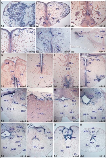
miR-9 expression in the adult zebrafish brain.
miR-9 is expressed in both periventricular and adjacent cells of proliferative zones and differentiating cells arising from these domains. miR-9 expressing cells are widespread in all brain subdivisions (fore-, mid- and hind-brain) although some areas such as the larval epithalamus and the hypothalamic lateral torus are devoid of expression (Table A). In addition to the brain, miR-9 is expressed in the inner retinal nuclear layer and ciliary marginal zone. miR-9 expression is highly conserved between larval and adult brain and we find only minor differences at the regional level (Table A,F). miR-9 is expressed in a few adult habenular and hypothalamic lateral torus cells, areas devoid of larval expression.

A. transverse section through the adult olfactory bulb showing miR-9 expressing cells in the internal (ICL), external (ECL) cellular and glomerular (GL) olfactory layers.
B. transverse section through the adult telencephalon showing miR-9 expressing cells in the ventral (Vv), lateral (Vl), central (Vc) and dorsal (Vd) nuclei of the ventral telencephalon (subpallium), medial (Dm), dorsal (Dd), central (Dc) and lateral (Dl) zones of the dorsal telencephalon (pallium).
C. higher magnification of B at the level of the subpallium showing miR-9 expressing cells in the ventral (Vv), central (Vc) and dorsal (Vd) nuclei of the ventral telencephalon.
D. higher magnification of B at the level of the pallium and dorsal subpallium showing miR-9 expressing cells in the dorsal nucleus of the ventral telencephalon (Vd), medial (Dm) and dorsal (Dd) zones of the dorsal telencephalon. Expression is present in many cells lining the ventricles in addition to presumptive neurons.
E. transverse section through the adult ventral telencephalon (caudal to sections B, C) showing periventricular (arrowheads) and lateral to the ventricle (arrows) miR-9 expressing cells in the ventral (Vv) and dorsal (Vd) nuclei of the ventral telencephalon.
F. transverse section through the adult hypothalamus showing miR-9 expressing cells in the dorsal zone of the periventricular hypothalamus (Hd). Arrowheads point at periventricular hypothalamic miR-9 expressing cells at the level of the lateral ventricular recess (lr) and more medially.
G. transverse section through the adult dorsal diencephalon showing a few miR-9 expressing cells in the dorsal habenular nucleus (Had), anterior (A) and ventromedial (VM) thalamic nuclei. Arrowheads point at periventricular miR-9 expressing cells.
H. transverse section through the adult caudal preoptic area at the level of the postoptic commissure (poc) showing miR-9 expressing cells in the posterior preoptic parvocellular nucleus (PPp). Blue arrowheads point at periventricular cells and black arrowheads point at miR-9 expressing cells at the level of the poc.
I. transverse section through the adult caudal preoptic area at the level of the postoptic commissure (poc) showing miR-9 expressing cells in the posterior parvocellular preoptic nucleus (PPp) and ventral zone of the periventricular hypothalamus (Hv).
J. transverse section through the adult diencephalon at the level of the posterior commissure (PC) showing miR-9 expressing cells in the ventral nucleus of the periventricular pretectum (PPv), dorsal (DP) and central (CP) posterior thalamic nuclei, periventricular nucleus of posterior tuberculum (TPp), ventral zone of the periventricular hypothalamus (Hv), anterior (PGa) and lateral (PGl) preglomerular nuclei.
K. transverse section through the adult caudal midbrain and rostral hindbrain showing miR-9 expressing cells in the perilemniscal nucleus (PL), nucleus of the lateral valvula (NLV), medial division of cerebellar valvula (Vam) and periventricular gray (pgz) and central (cz) zones of the optic tectum.
L. transverse section through the adult cerebellum showing miR-9 expressing cells in the granular (CeGL), ganglionic (CeGaL) and molecular (CeML) cerebellar layers.
M. transverse section through the adult rostral hindbrain showing miR-9 expressing cells in the superior reticular formation (SRF), superior raphe (SR), central gray (GC), nucleus of the lateral valvula (NLV) and granular cerebellar layer (CeGL).
N. transverse section through the adult hindbrain at the level of the octaval nerve (VIII) showing miR-9 expressing cells in the intermediate reticular formation (IMRF), anterior octaval nucleus (AON), central gray (GC), granular (CeGL) and molecular (CeML) cerebellar layers.
O. transverse section through the adult rostral hindbrain at the level of the octaval nerve (VIII, caudal to section N) showing miR-9 expressing cells in the intermediate reticular formation (IMRF), anterior (AON), secondary octaval (SO) and medial octavolateral (MON) nuclei, cerebellar crest (CC), granular (CeGL) and molecular (CeML) cerebellar layers.
P. transverse section through the adult hindbrain at the level of octaval nerve (VIII, caudal to section N) showing miR-9 expressing cells in the inferior reticular formation (IRF), descending octaval (DON) and caudal octavolateral (CON) nuclei and facial lobe (LVII). The arrowhead points at periventricular cells around the rhombencephalic ventricle expressing miR-9.
Q. transverse section through the adult caudal hindbrain at the level of the vagal motor nucleus (NXm) showing miR-9 expressing cells surrounding the rhombencephalic ventricle (rv), in the facial (LVII) and vagal (LX) lobes and inferior reticular formation (IRF).
R. transverse section through the adult caudal hindbrain at the level of the area postrema (AP) showing miR-9 expressing cells in the lateral part of the commissural nucleus of Cajal (NC) and surrounding (arrowheads) the rhombencephalic ventricle and medial longitudinal fascicle (MLF).

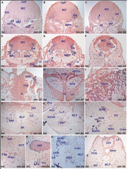
miR-34 expression in the zebrafish brain.
miR-34 shows very restricted expression in the larval brain with transcription restricted to ventral and lateral larval hindbrain areas, miR-34 conserves its expression in the adult hindbrain but additionally shows de novo expression in rostral and caudal nuclei, and thus changes dramatically its expression.
Specifically, at 3dpf and 5dpf, miR-34 is expressed in ventral and lateral medulla oblongata cells, (presumptive octaval area), locus coeruleus, trigeminal motor nuclei cells, Mauthner, and reticular formation cells (Table D). In the adult brain, miR-34 conserves its expression in respective nuclei and expands in specific nuclei and distinct cells of rostral brain, including epithalamic, pretectal, posterior tuberculum, tectal and tegmental cells, as well as in areas of dorsal hindbrain (cerebellar granular layer, facial and vagal lobes, Table I).

A. transverse section through the larval rostral hindbrain at the level of the octaval ganglion (OG) showing miR-34 expressing cells in the ventral medulla oblongata (MO), superior reticular formation (SRF), likely the trigeminal motor nucleus (NV?) and octaval ganglion (OG).
B. transverse section through the larval hindbrain at the level of the otic capsule (ot, caudal to section A) showing miR-34 expressing cells in the ventrolateral medulla oblongata (MO, likely octaval area, OA) and intermediate reticular formation (IMRF).
C. transverse section through the caudal larval hindbrain at the level of the inferior olive (IO) showing miR-34 expressing cells in the ventrolateral medulla oblongata (MO, arrowhead).
D. transverse section through the larval hindbrain at the level of the octaval ganglion (OG) showing miR-34 expressing cells in the ventrolateral medulla oblongata (MO, likely octaval area, OA), intermediate reticular formation (IMRF), Mauthner neuron (MAC) and octaval ganglion (OG).
E. transverse section through the larval hindbrain at the level of the octaval ganglion (OG, section caudal to section D) showing miR-34 expressing cells in the ventrolateral medulla oblongata (MO), likely octaval area (OA), intermediate reticular formation (IMRF) and octaval ganglion (OG). The arrowhead points at miR-34 expressing cells in the ventral hindbrain in proximity to the octaval nerve (VIII).
F. transverse section through the caudal larval hindbrain at the level of the inferior olive (IO) showing miR-34 expressing cells (arrowhead) in the ventrolateral medulla oblongata (MO), likely ventral to the vagal motor nucleus (NX).
G. transverse section through the adult epithalamus showing miR-34 expressing cells (arrowhead) in the dorsal habenular nucleus (Had).
H. transverse section through the adult hypothalamus showing miR-34 expressing cells in the nucleus of the paraventricular organ (nPVO).
I. transverse section through the adult cerebellum showing miR-34 expressing cells in the cerebellar granular layer (CeGL).
J. transverse section through the adult rostral hindbrain showing miR-34 expressing cells in the locus coeruleus (LC) and superior reticular formation (SRF).
K. transverse section through the adult hindbrain at the level of the trigeminal root showing miR-34 expressing cells in the primary sensory trigeminal nucleus (NVs), dorsal part of trigeminal motor nucleus (NVmd) and superior reticular formation (SRF).
L. transverse section through the adult lateral hindbrain at the level of the Mauhtner neuron (MAC) showing miR-34 expressing cells in the MAC, magnocellular (MaON), anterior (AON) octaval and medial octavolateral (MON) nuclei.
M. transverse section through the adult superficial pretectum showing miR-34 expressing cells in the magnocellular superficial pretectal nucleus (PSm) and the optic tectum (TeO).
N. transverse section through the adult pretectum and optic tectum showing miR-34 expressing cells in the tectal periventricular grey (pgz) and central (arrowhead) zones and accessory pretectal nucleus (APN).
O. transverse section through the adult lateral hindbrain at the level of the octaval nerve (VIII, caudal to section L) showing miR-34 expressing cells in the descending octaval (DON) and medial octavolateral (MON) areas and the intermediate reticular formation (IMRF). The arrowhead points at miR-34 expressing cells in the ventral hindbrain in proximity to the octaval nerve (VIII).
P. transverse section through the adult lateral hindbrain at the level of the vagal nerve (X), showing miR-34 expressing cells in the facial lobe (LVII), posterior octaval area (PON) and lateral to the medial longitudinal fascicle (MLF).

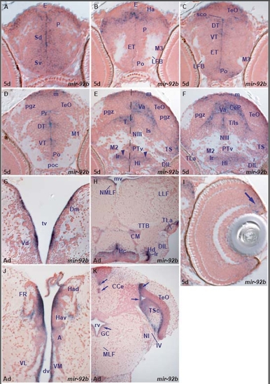
miR-92b expression in the zebrafish brain.
miR-92b is predominantly expressed in periventricular and adjacent cells corresponding to proliferative zones throughout the 5dpf larval brain (table A). It is also expressed in the retinal ciliary marginal zone. miR-92b expression is conserved in the same cell types in the adult brain (table F).

A. transverse section through the larval telencephalon showing miR-92b expressing cells in the ventral (Sv) and dorsal (Sd) subpallium, pallium (P) and epiphysis (E).
B. transverse section through the caudal telencephalon and rostral diencephalon showing miR-92b strongly expressing cells lining and close to the ventricle between the pallium (P) and epithalamus (habenula-Ha and epiphysis-E).
C. transverse section through the larval diencephalon and rostral optic tectum showing miR-92b expressing cells lining and close to the ventricle in the preoptic area, (Po), eminentia thalami (ET), ventral thalamus (VT), dorsal thalamus (DT) and the optic tectum (TeO).
D. transverse section through the larval diencephalon and optic tectum showing miR-92b expressing cells lining and close to the ventricle in the preoptic area (Po), ventral thalamus (VT), dorsal thalamus (DT), periventricular pretectum (Pr) and the proliferative zone of the optic tectum (m).
E. oblique transverse section through the larval midbrain, hypothalamus and rostral isthmus showing miR-92b expressing cells in the intermediate hypothalamus (Hi) around the lateral ventricular recess (lr, arrowheads), ventral posterior periventricular tuberculum (PTv), lining and close to the ventricle of tegmentum (T) and semicircular torus (TS), in the cerebellar valvula (Va) and proliferative zone of the optic tectum (m).
F. transverse section through the larval midbrain, hypothalamus and rostral isthmus showing miR-92b expressing cells in the intermediate hypothalamus (Hi) around the lateral ventricular ventricular recess (lr), lining and close to the ventricle in the tegmentum and isthmus (T/Is), semicircular torus (TS), in the cerebellar valvula (Va), cerebellar plate (CeP) and proliferative zone of the optic tectum (m).
G. transverse section through the adult telencephalon showing miR-92b expressing cells lining the telencephalic ventricle (tv) in the subpallium (dorsal nucleus of the ventral telencephalon,Vd) and the pallium (medial nucleus of the dorsal telencephalic area, Dm).
H. transverse section through the adult midbrain and hypothalamus showing miR-92b expressing cells lining/close to the ventricle in the dorsal (Hd-lr) and caudal (Hc) zones of the periventricular hypothalamus, mammillary body (CM) and lateral torus (TLa).
I. transverse section through the larval retina showing miR-92b expressing cells in the ciliary marginal zone (arrow).
J. transverse section through the adult dorsal diencephalon showing miR-92b expressing cells lining/close to the ventricle in the dorsal (Had) and ventral (Hav) habenular nuclei, anterior (A) and ventromedial (VM) thalamic nuclei.
K. transverse section through the adult cerebellum, isthmus and caudal midbrain. Arrows point at miR-92b expressing cells lining/close to the ventricle in the central gray (GC), optic tectum (TeO), central semicircular torus (TSc) and cerebellum (CCe).

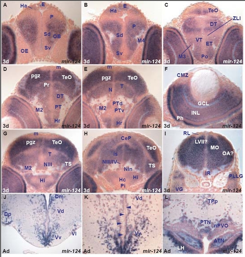
miR-124 expression in the zebrafish brain
miR-124 is expressed in virtually all differentiating cells throughout the 3dpf, 5dpf brain and retina and conserved in similar domains in the adult brain (Table B, G).

A. transverse section through the larval telencephalon and rostral epithalamus showing miR-124 expressing cells in the ventral (Sv) and dorsal (Sd) subpallium, pallium (P), olfactory bulb (OB) and habenula (Ha). Expression is absent from ventricular and periventricular cells.
B. transverse section through the larval caudal telencephalon and epithalamus showing miR-124 expressing cells in the ventral (Sv) and dorsal (Sd) subpallium, pallium (P), migrated telencephalic area (M4), habenula (Ha) and epiphysis (E).
C. transverse section through the larval diencephalon and rostral optic tectum showing miR-124 expressing cells in the preoptic area, (Po), eminentia thalami (ET), migrated eminentia thalami (M3), ventral thalamus (VT), dorsal thalamus (DT) and the optic tectum (TeO).
D. transverse section through the larval diencephalon and midbrain showing miR-124 expressing cells in the rostral hypothalamus (Hr), periventricular posterior tubercular area (PT), dorsal thalamus (DT), periventricular pretectum (Pr) and periventricular gray zone (pgz) of the optic tectum (TeO).
E. transverse section through the larval diencephalon and midbrain showing miR-124 expressing cells in the rostral hypothalamus (Hr), periventricular (dorsal-PTd, ventral-PTv) and migrated (M2) posterior tubercular area, tegmental area (T, at the level of the nucleus of medial longitudinal fascicle, N) and periventricular gray zone (pgz) of the optic tectum (TeO).
F. transverse section through the larval retina (dorsal to the right) showing miR-124 expressing cells in the photoreceptor (Ph), inner nuclear (INL) and ganglion cell (GCL) layers. Expression is absent from the ciliary marginal zone (CMZ).
G. transverse section through the larval hypothalamus and midbrain showing miR-124 expressing cells in the intermediate hypothalamus (Hi), migrated posterior tubercular area (M2), tegmental area (T, at the level of the oculomotor nucleus NIII), semicircular torus (TS) and periventricular gray zone (pgz) of the optic tectum (TeO).
H. transverse section through the larval hypothalamus, midbrain and isthmus showing miR-124 expressing cells in the intermediate hypothalamus (Hi), isthmic area (Is), cerebellar plate (CeP), semicircular torus (TS) and optic tectum (TeO).
I. transverse section through the hindbrain at the level of the posterior lateral line (PLLG) and vagal (VG) ganglia showing miR-124 expressing cells in the medulla oblongata (MO) and inferior raphe (IR).
J. transverse section through the adult telencephalon showing miR-124 expressing cells in the subpallium (ventral nucleus of the ventral telencephalon,Vv, lateral nucleus of the ventral telencephalon, Vl, dorsal nucleus of the ventral telencephalon,Vd) and the pallium (medial zone of the dorsal telencephalic area, Dm, posterior zone of the dorsal telencephalic area, Dp).
K. higher magnification of J at the level of ventral (Vv) and dorsal (Vd) nuclei of the ventral telencephalon, showing that miR-124 expressing cells are lateral to the ventricle (blue) whereas cells lining the ventricle are devoid of labelling (red, arrowheads).
L. transverse section through the adult hypothalamus showing miR-124 expressing cells in the lateral hypothalamic nucleus (LH), anterior tuberal nucleus (ATN), nucleus of the paraventricular organ (nPVO), posterior tuberal nucleus (PTN) and nucleus of posterior periventricular tuberculum (TPp).

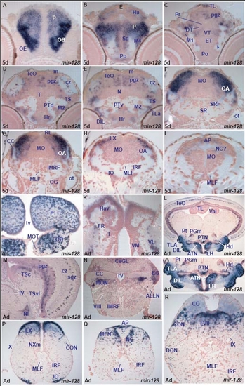
miR-128 expression in the zebrafish brain.
miR-128 shows spatially localized, conserved expression in specific larval and adult brain nuclei or cells. In addition, miR-128 expression shows major quantitative differences in levels of expression between structures in the larval brain.
At 5dpf, miR-128 is expressed in the entire olfactory bulb, pallium, and lateral medulla oblongata. In these regions, it is upregulated in the olfactory bulb, caudal pallial and lateral medulla oblongata cells. It is also expressed in distinct cells in the habenula, ventral thalamus, posterior tuberculum, lateral hypothalamus, periventricular and central tectal zones, semicircular torus and area postrema. It conserves its expression in these areas in the adult brain and given the good overall regional similarities in miR-128 expression between 5dpf and adult brain we suggest that the 5dpf expression in the medulla oblongata corresponds to cells of the presumptive octaval area, facial and vagal lobes, area postrema and commissural nucleus of Cajal. One difference in the expression pattern of miR-128 between larval and adult brain is expression in the adult inferior olive (Tables C, H).

A. transverse section through the rostral larval telencephalon showing miR-128 expressing cells in the olfactory bulb (OB) and pallium (P).
B. transverse section through the caudal telencephalon and epithalamus showing miR-128 expressing cells in the preoptic area (Po), dorsal subpallium (Sd), migrated telencephalic area (M4), pallium (P) and habenula (Ha).
C. transverse section through the larval diencephalon and optic tectum showing miR-128 expressing cells in the lateral eminentia thalami (ET, arrowhead), ventral thalamus (VT), migrated pretectal area (M1), periventricular gray zone (pgz) and longitudinal torus (TL) of the optic tectum.
D. transverse section through the larval hypothalamus and midbrain showing miR-128 expressing cells in the rostral hypothalamus (Hr), migrated posterior tubercular area (M2), semicircular torus (TS), periventricular gray (pgz) and central zone (cz) of the optic tectum (TeO).
E. transverse section through the larval hypothalamus and midbrain showing miR-128 expressing cells in the rostral hypothalamus (Hr), lateral torus (TLa), ventral periventricular (PTv) and migrated posterior tubercular area (M2), semicircular torus (TS), periventricular gray (pgz) and central zone (cz) of the optic tectum (TeO).
F. transverse section through the larval hindbrain at the level of the otic capsule (ot) showing miR-128 expressing cells in the lateral medulla oblongata (MO), including the octaval area (OA).
G. transverse section through the larval hindbrain at the level of the octaval ganglion (OG) showing miR-128 expressing cells in the lateral medulla oblongata (MO), including the octaval area (OA).
H. transverse section through the larval hindbrain caudal to the posterior lateral line ganglion showing miR-128 expressing cells in the lateral medulla oblongata (MO) likely in the octaval area (OA) and vagal lobe (LX).
I. transverse section through the larval caudal medulla oblongata (MO) showing miR-128 expressing cells in the area postrema (AP) and likely the commissural nucleus of Cajal (NC).
J. transverse section through the rostral adult telencephalon showing miR-128 expressing cells in the dorsal telencephalon/pallium (P) and olfactory bulb (OB) with the exception of the area of the medial olfactory tract (MOT).
K. transverse section through the adult epithalamus and thalamus showing miR-128 in the dorsal part of the ventral habenula (Hav) and ventrolateral thalamic nucleus (VL).
L. transverse section through the adult hypothalamus and midbrain showing miR-128 expressing cells in the lateral hypothalamic nucleus (LH), anterior tuberal nucleus (ATN), dorsal zone of the periventricular hypothalamus (Hd), diffuse nucleus of the inferior hypothalamic lobe (DIL), lateral torus (TLa), posterior tuberal nucleus (PTN), medial preglomeral nucleus (PGm), posterior thalamic nucleus (Pt) and optic tectum (TeO). For higher magnifications of the hypothalamus and optic tectum, see pictures M and O.
M. transverse section through the adult midbrain and isthmus (caudal to section L) showing miR-128 expressing cells in the isthmic nucleus (NI), central nucleus of semicircular torus (TSc), periventricular gray (pgz) and superficial grey (sgz) zones of the optic tectum. The asterisk marks the proliferative tectal zone devoid of miR-128 expression.
N. transverse section through the adult hindbrain at the level of the octaval nerve (VIII) showing miR-128 expressing cells in the medial octavolateral nucleus (MON), cerebellar granular eminence (EG) and granular cerebellar layer (CeGL).
O. higher magnification of section L at the level of the adult hypothalamus showing miR-128 expressing cells in the lateral hypothalamic nucleus (LH), anterior tuberal nucleus (ATN), dorsal zone of the periventricular hypothalamus (Hd), diffuse nucleus of the inferior hypothalamic lobe (DIL), lateral torus (TLa), posterior tuberal nucleus (PTN), medial preglomeral nucleus (PGm) and posterior thalamic nucleus (Pt).
P. transverse section through the adult hindbrain at the level of the vagal nerve (X, section caudal to R) showing miR-128 expressing cells in the inferior olive (IO), caudal octavolateral nucleus (CON) and vagal lobe (LX).
Q. transverse section through the adult hindbrain at the junction with the spinal cord (section caudal to P) showing miR-128 expressing cells in the area postrema (AP), commissural nucleus of Cajal (NC) and medial funicular nucleus (MFN).
R. transverse section through the adult hindbrain at the level of the cerebellar crest (CC, section caudal to N) showing miR-128 expressing cells in the descending octaval nucleus (DON), caudal octavolateral nucleus (CON) and facial lobe (LVII).

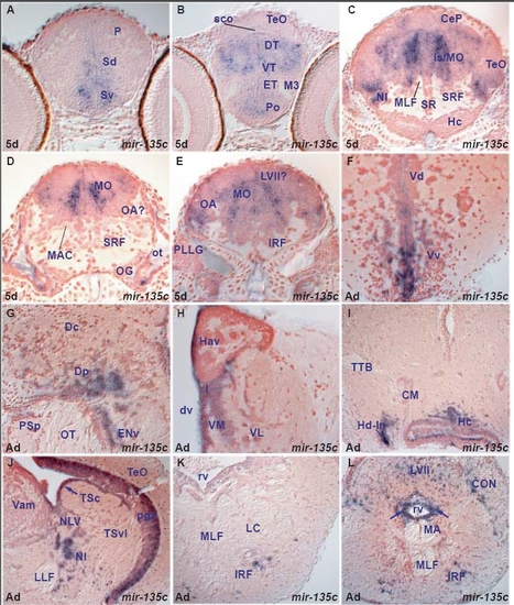
miR-135c expression in the zebrafish brain.
miR-135c is expressed in both periventricular and differentiating cells with a more restricted pattern compared to miR-9. miR-135c expression is mainly conserved between larval and adult brain but also shows some differences (Tables B,G). miR-135c expression is conserved in both larval and adult subpallium, caudal pallium, habenular, preoptic, thalamic, hypothalamic, pretectal, tegmental, cerebellar, isthmic and octaval nuclei. It is expressed in additional adult areas, devoid of larval expression, such as the caudal hypothalamus, periventricular and migrated posterior tuberculum nuclei, inferior raphe and reticular formation. In other hindbrain areas, it is difficult to draw conclusion about the conservation of miR-135c expression from larval to adult stages: miR-135c is robustly expressed in medial and central columns throughout the larval isthmic area and medulla oblongata but this is not the case in the adult. miR-135c is expressed in many cells of the adult facial and vagal lobes, and this expression may correspond to part of the larval one.

A. transverse section through the larval telencephalon showing miR-135c expressing cells in the ventral (Sv) and dorsal (Sd) subpallium.
B. transverse section through the larval diencephalon and rostral optic tectum showing miR-135c expressing cells in the preoptic area (Po), eminentia thalami (ET), ventral (VT) and dorsal thalamus (DT).
C. transverse section through the larval caudal midbrain, hypothalamus, cerebellum and isthmus/medulla oblongata showing miR-135c expressing cells in the isthmic nucleus (NI), cerebellar plate (CeP), optic tectum (TeO) and isthmic/medulla oblongata areas (Is/MO).
D. transverse section through the larval hindbrain at the level of the octaval ganglion (OG) showing miR-135c expressing cells in the medulla oblongata (MO).
E. transverse section through the larval hindbrain at the level of the posterior lateral line ganglion (PLLG) showing miR-135c expressing cells in the medulla oblongata (MO) likely including the presumptive octaval area (OA).
F. transverse section through the adult telencephalon showing miR-135c expressing cells lining or lateral to the telencephalic ventricle in the subpallium (ventral nucleus of the ventral telencephalon,Vv, dorsal nucleus of the ventral telencephalon,Vd).
G. transverse section through the adult caudal telencephalon showing miR-135c expressing cells in the posterior zone of the dorsal telencephalon (Dp) and ventral part of the entopeduncular nucleus (ENv).
H. transverse section through the adult dorsal diencephalon showing miR-135c expressing cells lining or close to the diencephalic ventricle (dv) in the ventral habenular nucleus (Hav), intermediate (I), ventromedial (VM) and ventrolalateral (VL) thalamic nuclei.
I. transverse section through the adult midbrain and hypothalamus showing miR-135c expressing cells lining/close to the ventricle in the dorsal (Hd-lr, around the lateral ventricular recess) and caudal (Hc) zones of the periventricular hypothalamus.
J. transverse section through the adult caudal midbrain and isthmus showing miR-135c expressing cells in the tectal periventricular gray zone (pgz), periventricular central semicircular torus (arrow, TSc) nucleus of lateral valvula (NLV) and isthmic nucleus (NI).
K. transverse section through the adult isthmus showing miR-135c expressing cells in the inferior reticular formation (IRF).
L. transverse section through the caudal medulla oblongata showing miR-135c expressing cells in the inferior reticular formation (IRF), caudal octavolateral nucleus (CON), facial lobe (LVII) and surrounding the rhombencephalic ventricle (rv, arrows).

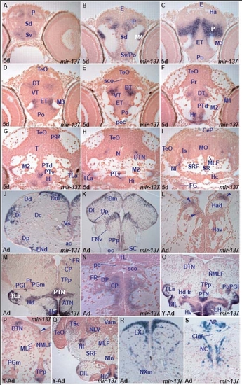
miR-137 expression in the zebrafish brain.
miR-137 shows spatially localized, conserved expression in specific larval and adult brain nuclei or cells (Table C, H). miR-137 expression is conserved in many larval and adult areas including the subpallium, ventro-medial and caudal pallium, preoptic area, dorsal thalamus, hypothalamus and ventral posterior tubercular area. Given the good correspondence of localized expression between larval and adult brains, this allows us to annotate specific nuclei in the larval tegmentum, isthmus and medulla oblongata. We suggest that the cells in the larval tegmentum and medulla oblongata correspond to the midbrain dorsal tegmental nucleus, lateral nucleus of cerebellar valvula, isthmic nucleus, facial and glossopharyngeal/vagal lobes, vagal motor nucleus and area postrema. Despite the well-conserved pattern of expression between larval and adult zebrafish brain, we observe minor differences. miR-137 is expressed in adult dorsal lateral habenular cells and a few tectal periventricular gray zone and migrated posterior tuberculum cells.

A. transverse section through the larval telencephalon showing miR-137 expressing cells in the ventral (Sv) and dorsal (Sd) subpallium and pallium (P).
B. transverse section through the caudal telencephalon and epithalamus showing miR-137 expressing cells in the ventral subpallium(Sv)/preoptic area (Po), dorsal subpallium (Sd), migrated telencephalic area (M4) and pallium (P).
C. transverse section through the caudal telencephalon and epithalamus showing miR-137 expressing cells in the pallium (P) and eminentia thalami (ET).
D. transverse section through the larval diencephalon and rostral optic tectum showing miR-137 expressing cells in the eminentia thalami (ET), ventral thalamus (VT) and dorsal thalamus (DT).
E. transverse section through the larval diencephalon and rostral optic tectum (caudal to section D) showing miR-137 expressing cells in the eminentia thalami (ET), ventral thalamus (VT) and dorsal thalamus (DT).
F. transverse section through the larval diencephalon and midbrain showing miR-137 expressing cells in the rostral hypothalamus (Hr), dorsal periventricular posterior tuberculum (PTd) and dorsal thalamus (DT).
G. transverse section through the larval diencephalon and midbrain showing miR-137 expressing cells in the intermediate hypothalamus (Hi), lateral hypothalamic torus (TLa) and ventral periventricular posterior tuberculum (PTv).
H. transverse section through the larval diencephalon and midbrain showing miR-137 expressing cells in the lateral hypothalamic torus (TLa), ventral periventricular posterior tuberculum (PTv) and midbrain dorsal tegmental nucleus (DTN).
I. transverse section through the larval caudal midbrain and rostral hindbrain at the level of the facial ganglion (FG) showing miR-137 expressing cells in the isthmic area (Is) including the isthmic nucleus (NI).
J. transverse section through the adult telencephalon at the level of the anterior commissure (ac) showing miR-137 expressing cells in the supracommissural nucleus of the ventral (subpallial) telencephalic area (Vs), posterior (Dp), lateral (Dl), dorsal (Dd), medial (Dm) zones of the dorsal (pallial) telencephalic area and the dorsal entopeduncular nucleus (ENd).
K. transverse section through the adult caudal telencephalon at the level of the optic chiasma (oc) showing miR-137 expressing cells in the posterior (Dp), lateral (Dl) and medial (Dm) zones of the dorsal (pallial) telencephalic area, posterior preoptic parvocellular (PPp) and suprachiasmatic (SC) nuclei.
L. transverse section through the adult epithalamus showing miR-137 expressing cells in the dorsal habenular nucleus (Had, arrowheads).
M. transverse section through the adult diencephalon showing miR-137 expressing cells in the ventral zone of the hypothalamus (Hv), anterior tuberal nucleus (ATN), dorsal zone of the periventricular hypothalamus (Hd), lateral torus (TLa), posterior tuberal nucleus (PTN), medial preglomeral nucleus (PGm), posterior thalamic nucleus (Pt) and central posterior thalamic nucleus (CP). For higher magnification of the thalamus, see section N.
N. Higher magnification of a transverse section (level caudal to section M) through the adult dorsal diencephalon, at the level of the posterior commissure (pc) showing miR-137 expressing cells in the central posterior thalamic nucleus (CP).
O. transverse section through the young adult diencephalon and midbrain showing miR-137 expressing cells in the ventral zone of the hypothalamus (Hv), lateral hypothalamic nucleus (LH), dorsal zone of the periventricular hypothalamus around the lateral hypothalamic ventricular recess (Hd-lr), diffuse nucleus of inferior lobe (DIL), lateral torus (TLa), posterior tuberal nucleus (PTN), ventral part of the nucleus of the posterior periventricular tuberculum (TPp), posterior thalamic nucleus/lateral preglomeral nucleus (Pt/PGl) and midbrain dorsal tegmental nucleus (DTN).
P. Higher magnification of the dorsal part of the transverse section P showing miR-137 expressing cells in the midbrain dorsal tegmental nucleus (DTN), ventral part of the nucleus of the posterior periventricular tuberculum (TPp) and a few cells in the proximity of the mesencephalic ventricle (arrowhead).
Q. transverse section through the young adult caudal hypothalamus, midbrain and isthmus showing miR-137 expressing cells in the caudal zone of the hypothalamus (Hc), diffuse nucleus of the hypothalamic inferior lobe (DIL), isthmic nucleus (NI), nucleus of lateral cerebellar valvula (NLV), central nucleus of semicircular torus (TSc) and optic tectum (TeO).
R. transverse section through the adult caudal hindbrain at the level of the vagal nerve showing miR-137 expressing cells in the vagal lobe (LX) and vagal motor nucleus (NXm).
S. transverse section through the adult hindbrain at the junction with the spinal cord (caudal to section R) showing miR-137 expressing cells in the area postrema (AP) and commissural nucleus of Cajal (NC).

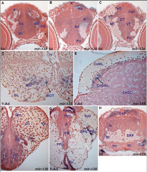
miR-138 expression in the zebrafish brain
miR-138 is expressed in differentiated cells with more restriction compared to miR-124. Expression is largely conserved between larval and adult brain but there are some differences (Tables B,G). With the exception of the medulla oblongata where miR-138 larval expression is widespread and thus difficult to correlate with the adult, we observe the following similarities and differences between larval and young adult expression: miR-138 is expressed in both larval and young adult brains in the olfactory bulb, pallial and subpallial areas, preoptic area, dorsolateral habenular cells, dorsal posterior thalamic nuclei, hypothalamic region, tectal cells, isthmic area and cerebellar cells. Furthermore it is expressed in the adult medial octavolateral nucleus, facial and vagal lobe and reticular formation. This adult hindbrain expression may partially correspond to the larval expression in lateral isthmus and central and medial part of the medulla oblongata. miR-138 is not expressed in the young adult ventral thalamus and semicircular torus whereas it is expressed in these areas at 5dpf. Conversely, it is expressed in adult migrated posterior tuberculum areas, migrated pretectal nuclei, longitudinal torus and cerebellar valvula cells, areas devoid of staining in the larval brain.

A. transverse section through the larval telencephalon showing miR-138 expressing cells in the ventral (Sv) and dorsal (Sd) subpallium, pallium (P) and olfactory bulb (OB).
B. oblique transverse section through the larval caudal telencephalon and epithalamus showing miR-138 expressing cells in the preoptic area (Po), dorsal subpallium (Sd), pallium (P) and habenula (Ha).
C. transverse section through the larval diencephalon and optic tectum showing miR-138 expressing cells in the ventral thalamus (VT), dorsal thalamus (DT) and the periventricular gray zone of the optic tectum (pgz).
D. transverse section through the adult olfactory bulb showing miR-138 expressing cells in the internal granular layer (ICL).
E. transverse section through the young adult cerebellum showing miR-138 expressing cells in the cerebellar ganglionic layer (CeGAL).
F. transverse section through the young adult telencephalon showing miR-138 expressing cells in the subpallium (ventral nucleus of the ventral telencephalon, Vv, dorsal nucleus of the ventral telencephalon,Vd) and the pallium (medial zone of the dorsal telencephalic area, Dm).
G. transverse section through the young adult diencephalon showing miR-138 expressing cells in the ventral periventricular zone of the hypothalamus (Hv), dorsal habenular nucleus (Had), parvocellular superficial (PSp) and central (CPN) pretectal nuclei.
H. transverse section through the larval isthmus, hypothalamus and caudal midbrain showing miR-138 expressing cells in the lateral hypothalamic torus (TLa), isthmic area (Is), and cerebellar plate (CeP).

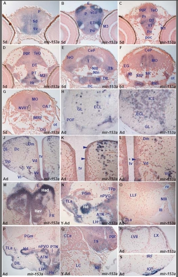
miR-153a expression in the zebrafish brain.
miR-153a shows expression in both periventricular and differentiating cells in the larval brain and has more restricted expression compared to miR-124. It is expressed in periventricular cells only in a few cases. In addition, miR-153a expression shows quantitative differences from one area to another and is conserved in many areas between larval and adult rostral brain but also presents some regional differences (Tables C,H).
Rostrally, miR-153a is expressed in both larval and adult telencephalic pallial and subpallial areas, preoptic area, habenula, dorsal and ventral thalamus, hypothalamus, posterior tuberculum, mesencephalic tectal periventricular gray zone and interpeduncular nucleus. In addition to these similarities between rostral larval and adult brain, we observe some differences: miR-153a is expressed only in the adult olfactory bulb whereas it is expressed only in the larval periventricular pretectum. Furthermore, it is expressed in the entire larval tegmentum but in the adult it is restricted in the semicircular torus. Caudally, miR-153a is expressed in the young adult and adult hindbrain, in the isthmic nucleus, nucleus of lateral valvula, perilemniscal nucleus, facial and vagal lobes and area postrema. Within the larval hindbrain we observe columns of miR-153a expression but it is difficult to establish a precise correlation with the adult nuclei. Finally, we observe quantitative differences in miR-153a expression between regions. For example miR-153a expression is particularly highly expressed in the larval habenula and migrated eminentia thalami.

A. transverse section through the larval telencephalon showing miR-153a in the ventral (Sv) and dorsal (Sd) subpallium and pallium (P).
B. transverse section through the larval caudal telencephalon and diencephalon showing miR-153a expressing cells in the preoptic area (Po), eminentia thalami (ET), migrated eminentia thalami (M3), pallium (P) and habenula (Ha).
C. transverse section through the larval diencephalon and rostral optic tectum showing miR-153a expressing cells in the preoptic area (Po), ventral thalamus (VT), dorsal thalamus (DT), periventricular pretectum (Pr) and periventricular gray zone (pgz) of the optic tectum (TeO).
D. transverse section through the larval diencephalon and midbrain showing miR-153a expressing cells in the rostral hypothalamus (Hr), periventricular (PT) and migrated (M2) posterior tubercular area, dorsal thalamus (DT) and periventricular gray zone (pgz) of the optic tectum (TeO).
E. transverse section through the larval caudal hypothalamus, midbrain and rostal hindbrain showing miR-153a expressing cells in the intermediate (Hi) and caudal (Hc) hypothalamus, diffuse nucleus of inferior lobe (DIL), interpeduncular nucleus (NIn), tegmentum (T), oculomotor nucleus (NIII), semicircular torus (TS) and optic tectum (TeO).
F. transverse section through the larval rostal hindbrain and caudal hypothalamus showing miR-153a expressing cells in the central medulla oblongata (MO), isthmic nucleus (NI), superior raphe (SR) and caudal hypothalamus (Hc).
G. transverse section through the larval hindbrain at the level of the octaval ganglion (OG) showing miR-153a expressing cells in the central medulla oblongata (MO).
H. transverse section through the rostral adult olfactory bulb showing miR-153a expressing cells in the glomerular (GL) and external cellular (ECL) layers.
I. transverse section through the adult olfactory bulb (caudal to section H) showing miR-153a expressing cells in the glomerular (GL), external (ECL) and internal (ICL) cellular layers.
J. transverse section through the adult telencephalon showing miR-153a expressing cells in the ventral (Vv), lateral (Vl), central (Vc) and dorsal (Vd) nuclei of the ventral telencephalon (subpallium), medial (Dm), central (Dc), lateral (Dl) and posterior (Dp) zones of the dorsal telencephalon (pallium).
K. higher magnification of J through the adult dorsal telencephalon showing strongly miR-153a expressing cells along the dorsal part of the telencephalic ventricle (tv, asterisk) and the lateral part of the medial zone of the dorsal telencephalon (Dm) and few weakly expressing miR-153a cells along the medial ventricular wall (arrowheads).
L. higher magnification of J through the adult medial telencephalic part, ventral to section K, showing strongly miR-153a expressing cells in the lateral part of Dm (medial zone of dorsal telencephalon), central nucleus of the ventral telencephalon (Vc) and weakly expressing cells along the medial ventricle in the dorsal nucleus of the ventral telencephalon (Vd, arrowhead).
M. transverse section through the adult epithalamus showing miR-153a expressing cells in the dorsal (Had) and ventral (Hav) habenular nuclei.
N. transverse section through the young adult hypothalamus showing miR-153a expressing cells in the lateral hypothalamic nucleus (LH), anterior tuberal nucleus (ATN), dorsal zone of the periventricular hypothalamus (Hd), diffuse nucleus of the inferior hypithalamic lobe (DIL), lateral torus (TLa), nucleus of the paraventricular organ (nPVO), medial preglomeral nucleus (PGm) and periventricular nucleus of posterior tuberculum (TPp).
O. transverse section through the adult tegmentum at the level of the oculomotor nucleus (NIII) showing miR-153a expression only in the lateral hypothalamic torus (TLa).
P. transverse section through the adult hypothalamus showing miR-153a expressing cells in the lateral hypothalamic nucleus (LH), anterior tuberal nucleus (ATN), ventral zone of the periventricular hypothalamus (Hv), dorsal zone of the periventricular hypothalamus (Hd), diffuse nucleus of the inferior hypothalamic lobe (DIL), lateral torus (TLa), nucleus of the paraventricular organ (nPVO) and posterior tuberal nucleus (PTN).
Q. transverse section through the young adult isthmus and caudal midbrain showing miR-153a expressing cells in the semicircular torus (TS), tectal periventricular gray zone (pgz) and istmic nucleus (NI).
R. transverse section through the adult caudal hindbrain showing miR-153a expressing cells in the facial (LVII) and vagal (LX) lobes.
S. transverse section through the adult caudal hindbrain (ventral to section Q) showing miR-153a expressing cells in the inferior olive (IO).

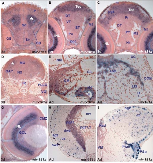
miR-181a expression in the zebrafish brain.
miR-181a and miR-181b belong to the same miRNA family and differ in three nucleotides located outside the seed region. These miRNAs show similar, spatially localised expression in the larval brain. miR-181a and miR-181b expression is quantitatively different from one area to another in the larval brain with strong expression in the retina and brain areas associated with the visual system. miR-181a and miR-181b expression is largely conserved to adult stage although there is downregulation in some areas. Despite overall conservation, we noticed subtle differences in the adult expression of miR-181a and miR-181b that were not obvious at larval stages.
In the larval brain, miR-181a and miR-181b are strongly expressed in retinal ganglion and inner cell layers, migrated pretectal, and tectal cells, that is, cells associated with vision. There are two more areas with strong miR-181a and miR-181b expression, a group of cells located in the central part of the pallium, close to the olfactory bulb and dorsal subpallium, and one in the central medulla oblongata whereas the rest of the larval brain contains weakly expressing cells. Given the adult expression in the olfactory bulb and facial and vagal lobes, the strong telencephalic and medulla oblongata larval expression may correspond to cells that populate these areas. While comparing the regional expression of miR-181a and miR-181b, significant regional differences are not obvious at larval stages (Table C).
Overall, miR-181a and miR-181b conserve their regional expression between larval and adult brain (Table H). For example, miR-181a and miR-181b are expressed in adult pretectal, tectal, hypothalamic and cerebellar cells as it is the case in the larval brain. But there are also differences between larval and adult expression. miR-181a and miR-181b expression is downregulated in the adult thalamus, periventricular posterior tuberculum and tegmentum. In addition, quantitative differences of expression observed in the larval brain are absent from the adult brain.
Finally, comparison of adult miR-181a and miR-181b expression reveals similar regional expression but differences at the cellular level within some areas. Good examples are the caudal hypothalamus and the facial lobe. Both miRNAs are expressed in these areas but likely in different cells. In other areas, like pretectum and tectum, telencephalon and olfactory bulb, they are likely expressed in the same cell types.

A. transverse section through the larval telencephalon showing miR-181a expressing cells throughout the ventral (Sv) and dorsal (Sd) subpallium, pallium (P) and olfactory bulb (OB). The asterisk marks the strongly expressing miR-181a cell group in the central pallium, close to OB and Sd.
B. transverse section through the larval diencephalon and optic tectum (TeO) showing strongly miR-181a expressing cells in the TeO and migrated pretectal area (M1) and weakly in the preoptic area (Po), ventral thalamus (VT), dorsal thalamus (DT) and eminentia thalami (ET).
C. transverse section through the larval diencephalon and optic tectum (TeO) showing strongly miR-181a expressing cells in the TeO and migrated pretectal area (M1) and weakly in the rostral hypothalamus (Hr), periventricular (PT) and migrated (M2) posterior tuberculum, dorsal thalamus (DT) and periventricular pretectum (Pr).
D. transverse section through the larval hindbrain at the level of the posterior lateral line (PLLG) and vagal (VG) ganglia showing miR-181a expressing cells in the medulla oblongata (MO). The asterisk marks the strongly miR-181a expressing cells in the lateral medulla oblongata likely in proximity to the octaval area (OA?).
E. transverse section through the adult hypothalamus showing miR-181a expressing cells in the caudal part of the dorsal zone of the periventricular hypothalamus (Hd) around the ventricular recess (lr) and mammillary body (CM).
F. transverse section through the adult caudal hindbrain showing miR-181a expressing cells in the facial (LVII) and glossopharyngeal (LIX) lobes.
G. transverse section through the larval retina showing miR-181a expressing cells in the inner part (amacrine cells) of the inner nuclear layer (INL) and the ganglion cell layer (GCL).
H. transverse section through the adult optic tectum showing miR-181a expressing cells in the superficial gray zone (sgz), central zone (cz) and the third division of the periventricular gray zone (pgz3).
I. transverse section through the adult dorsal diencephalon and rostral optic tectum showing miR-181a expressing cells in the superficial gray zone (sgz), central zone (cz), parvocellular (PSp) and magnocellular (PSm) superficial pretectal nuclei.

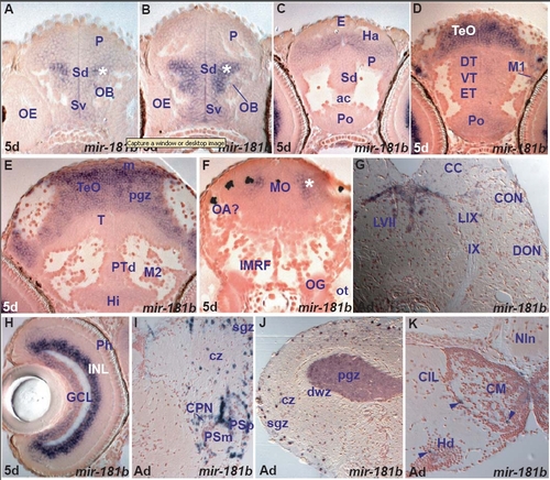
miR-181b expression in the zebrafish brain.
miR-181a and miR-181b belong to the same miRNA family and differ in three nucleotides located outside the seed region. These miRNAs show similar, spatially localised expression in the larval brain. miR-181a and miR-181b expression is quantitatively different from one area to another in the larval brain with strong expression in the retina and brain areas associated with the visual system. miR-181a and miR-181b expression is largely conserved to adult stage although there is downregulation in some areas. Despite overall conservation, we noticed subtle differences in the adult expression of miR-181a and miR-181b that were not obvious at larval stages.
In the larval brain, miR-181a and miR-181b are strongly expressed in retinal ganglion and inner cell layers, migrated pretectal, and tectal cells, that is, cells associated with vision. There are two more areas with strong miR-181a and miR-181b expression, a group of cells located in the central part of the pallium, close to the olfactory bulb and dorsal subpallium, and one in the central medulla oblongata whereas the rest of the larval brain contains weakly expressing cells. Given the adult expression in the olfactory bulb and facial and vagal lobes, the strong telencephalic and medulla oblongata larval expression may correspond to cells that populate these areas. While comparing the regional expression of miR-181a and miR-181b, significant regional differences are not obvious at larval stages (Table C).
Overall, miR-181a and miR-181b conserve their regional expression between larval and adult brain (Table H). For example, miR-181a and miR-181b are expressed in adult pretectal, tectal, hypothalamic and cerebellar cells as it is the case in the larval brain. But there are also differences between larval and adult expression. miR-181a and miR-181b expression is downregulated in the adult thalamus, periventricular posterior tuberculum and tegmentum. In addition, quantitative differences of expression observed in the larval brain are absent from the adult brain.
Finally, comparison of adult miR-181a and miR-181b expression reveals similar regional expression but differences at the cellular level within some areas. Good examples are the caudal hypothalamus and the facial lobe. Both miRNAs are expressed in these areas but likely in different cells. In other areas, like pretectum and tectum, telencephalon and olfactory bulb, they are likely expressed in the same cell types.

A. transverse section through the larval telencephalon showing miR-181b expressing cells throughout the ventral (Sv) and dorsal (Sd) subpallium, pallium (P) and olfactory bulb (OB). The asterisk marks the strongly expressing miR-181b cell group in the central pallium, close to the OB and Sd.
B. transverse section through the larval telencephalon (caudal to section A) showing miR-181b expressing cells throughout the ventral (Sv) and dorsal (Sd) subpallium, pallium (P) and olfactory bulb (OB). The asterisk marks the strongly expressing miR-181b cell group in the central pallium, close to the OB and Sd.
C. transverse section through the larval caudal telencephalon at the level of the anterior commissure (ac) showing miR-181b expressing cells in the preoptic area (Po), dorsal subpallium (Sd), pallium (P) and habenula (Ha).
D. transverse section through the larval diencephalon and optic tectum (TeO) showing miR-181b expressing cells in the TeO, preoptic area (Po), eminentia thalami (ET), ventral (VT) and dorsal (DT) thalamus and migrated pretectal area (M1).
E. transverse section through the larval hypothalamus and midbrain showing strongly expressing miR-181b cells in the optic tectum (TeO, periventricular gray zone-pgz and proliferative zone-m).
F. transverse section through the larval hindbrain at the level of the otic capsule (ot) showing miR-181b expressing cells in the medulla oblongata (MO). The asterisk marks the strongly miR-181b expressing cells in the central medulla oblongata likely in proximity to the octaval area (OA?).
G. transverse section through the adult caudal hindbrain showing miR-181b expressing cells in the facial lobe (LVII) and caudal octavolateral nucleus (CON).
H. transverse section through the larval retina showing miR-181b expressing cells in the inner part (amacrine cells) of the inner nuclear layer (INL) and the ganglion cell layer (GCL).
I. transverse section through the adult dorsal diencephalon and rostral optic tectum showing miR-181b expressing cells in the tectal superficial gray (sgz) and central (cz), zones, parvocellular (PSp), magnocellular (PSm) and central (CPN) superficial pretectal nuclei.
J. transverse section through the adult optic tectum showing miR-181b expressing cells in the superficial gray (sgz), central (cz) and periventricular gray (pgz) zones.
K. transverse section through the adult hypothalamus showing miR-181b expressing cells (arrowheads) in the caudal part of the dorsal zone of the periventricular hypothalamus around the lateral ventricular recess (Hd-lr) and mamillary body (CM).

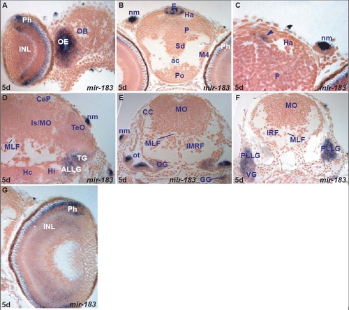
miR-183 expression in the zebrafish brain.
miR-183 expression is cell type specific in larvae. It is expressed in retinal photoreceptors and weakly in some inner nuclear layer cells, pineal cells that are again likely to be photoreceptors and perhaps also in parapineal photoreceptors. Outside of the CNS, miR-183 is expressed in cells that include peripheral sensory neuromasts, olfactory sensory neurons and hair cells of the ear. miR-182 and miR-96 show almost identical expression patterns to miR-183 (Table E) although expression is not as robust.

A. transverse section through the larval rostral brain and retina showing miR-183 expressing cells in the olfactory epithelium (OE), retinal photoreceptor (Ph) and inner nuclear (INL) layers.
B. transverse section through the larval telencephalon and rostral epithalamus showing miR-183 expressing cells in the epiphysis (E) and neuromasts (nm).
C. transverse section through the larval epithalamus and pallium (P) showing miR-183 expressing cells medial to the habenula (Ha) likely corresponding to parapineal organ (arrowhead) and neuromasts (nm).
D. transverse section through the larval midbrain and isthmus showing miR-183 expressing cells in the trigeminal (TG), anterior lateral line (ALLG) ganglia and neuromasts (nm).
E. transverse section through the larval hindbrain showing miR-183 expressing cells in the otic capsule (ot), octaval (OG) and glossopharyngeal (GG) ganglia and neuromasts (nm).
F. transverse section through the larval caudal hindbrain showing miR-183 expressing cells in the posterior lateral line (PLLG) and vagal (VG) ganglia.
G. transverse section through the larval retina showing miR-183 expressing cells in the retinal photoreceptor (Ph) and inner cellular layer (INL).

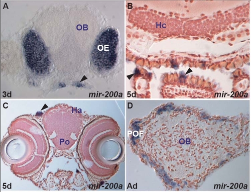
miR-200a expression in the zebrafish brain.
miR-200a shows cell-type specific expression (Table E). It is expressed in sensory cells of the olfactory epithelium, taste buds, ear, neuromasts as well as the adult primary olfactory fiber layer.

A. transverse section through the rostral part of the larval head showing miR-200a expressing cells in the olfactory epithelium (OE) and the taste buds (arrowhead).
B. transverse section through the caudal part of the larval head showing miR-200a expressing cells in the taste buds (arrowheads).
C. transverse section through the larval part of the head at the level of epithalamus (Ha, habenula) showing miR-200a expressing cells in neuromasts (arrowhead).
D. transverse section through the adult olfactory bulb (OB) showing miR-200a expressing cells in the primary olfactory fiber layer (POF). These may be transcripts in axons of olfactory sensory neurons.

miR-218a expression in the zebrafish brain.
miR-218a shows cell type specific expression in cranial motor nuclei (III, V, VI, VII, IX, X) and spinal motoneurons (Table E). miR-218a expression is conserved in adult motor nuclei, but also expands in rostral brain areas including the ventral telencephalon, magnocellular preoptic area, ventral and lateral hypothalamic nuclei, optic tectum and inferior olive (Table I).

A. transverse section through the larval hindbrain at the level of the octaval ganglion (OG) showing miR-218a expression in the trigeminal motor nucleus (NV).
B. transverse section through the larval hindbrain at the level of the otic capsule (ot, caudal to section A) showing miR-218a expression in the facial (NVII) and abducens (NVI) motor nuclei.
C. transverse section through the larval hindbrain at the level of the otic capsule (ot, caudal to section B) showing miR-218a expression in the facial motor nucleus (NVII).
D. transverse section through the larval hindbrain at the level of the posterior lateral line (PLLG) and vagal (VG) ganglia showing miR-218a expression in the glossopharyngeal motor nucleus (NIX).
E. transverse section through the larval caudal hindbrain (caudal to section D) showing miR-218a expression in the vagal motor nucleus (NX).
F. transverse section through the larval caudal hindbrain (caudal to section E) showing miR-218a expressing cells in the vagal motor nucleus (NX) and motor neurons (MN) of the caudal inferior reticular formation.
G. parasagittal confocal section through the larval hindbrain showing miR-218a expressing cells in the facial (NVII) and vagal (NX) motor nuclei.
H. parasagittal confocal section through the larval hindbrain showing cells expressing Tg(isl1:gfp) in the facial (NVII) and vagal (NX) motor nuclei.
I. superimposition of sections G and H showing co-localization (in yellow) of miR-218a (red) and GFP (green) expression in the facial (NVII) and vagal (NX) motor nuclei.
J. transverse section through the larval spinal cord showing miR-218a expressing cells in motor neurons (MN).
K. transverse section through the adult preoptic area at the level of the optic chiasma (oc) showing miR-218a expressing cells in the magnocellular preoptic nucleus (PM).
L. transverse section through the adult ventral hypothalamus showing miR-218a expressing cells in the lateral hypothalamic nucleus (LH) and ventral zone of the periventricular hypothalamus (Hv).
M. transverse section through the adult caudal midbrain showing weak miR-218a expressing cells in the oculomotor nucleus (NIII).
N. transverse section through the adult hindbrain at the level of the trigeminal nerve (ventral motor root, Vmv) showing miR-218a expressing cells in the ventral part of the trigeminal motor nucleus (NVmv).
O. transverse section through the adult hindbrain at the level of the octaval nerve (VIII) showing miR-218a expressing cells in the rostral part of the abducens nucleus (NVIr).
P. transverse section through the adult hindbrain at the level of the octaval nerve (VIII, caudal to section O) showing miR-218a expressing cells in the caudal part of the abducens (NVIc) and motor facial (NVIIm) nuclei.
Q. transverse section through the adult caudal hindbrain at the level of the vagal nerve (caudal to section P) showing miR-218a expressing cells in the vagal motor nucleus (NXm) and inferior olive (IO).
R. transverse section through the adult spinal cord showing miR-218a expressing cells in motor neurons (MN) of the ventral horn (VH).

miR-219 expression in the zebrafish brain.
miR-219 is expressed in both periventricular and differentiated cells in the larval brain and has more restricted expression in differentiating cells compared to miR-124. Transcripts are localized in periventricular and adjacent cells only in few cases. miR-219 shows pronounced changes between larval and adult brain expression patterns. miR-219 transcript localization suggests downregulation of expression during maturity and conserved or de novo expression in distinct cells, possibly glia.
In the 5dpf larval brain, miR-219 is expressed in the entire ventral and dorsal thalamus, tegmentum, isthmus, medulla oblongata and ventral hindbrain areas such as the reticular formation. In addition, it is expressed in most pretectal cells, in a few pallial, ventral habenular, dorsal posterior tubercular, tectal and lateral cerebellar plate cells. It is absent from the caudal preoptic area, hypothalamus, retina and Mauthner cells (Table B). In the adult brain, it is widely expressed only in the caudal hindbrain. In the rostral adult brain, expression is often downregulated as is the case in the ventral thalamus and is limited to few distinct cells found in areas that are positive (pallium, epithalamus, central posterior (dorsal) thalamic nucleus, posterior tuberculum, pretectum, tectum, tegmentum, cerebellum) or negative in the larval brain (olfactory bulb, subpallium, caudal preoptic area, hypothalamus). In some cases these distinct adult cells, possibly glia, are associated with nerves or tracts such as the lateral olfactory tract, optic tract or commissures such as anterior, postoptic and tectal commissure, whereas the equivalent pathways in larvae are devoid of staining (Table G).

A. transverse section through the larval caudal telencephalon and rostral diencephalon showing miR-219 weakly expressing cells in the rostral preoptic area (Po), eminentia thalami (ET), pallium (P), dorsal thalamus (DT) and habenula (Ha).
B. transverse section through the larval diencephalon and optic tectum showing miR-219 expressing cells in the ventral (VT) and dorsal (DT) thalamus and pretectum (Pr).
C. transverse section through the larval diencephalon and optic tectum showing miR-219 expressing cells in the dorsal periventricular posterior tuberculum (PTd), dorsal thalamus (DT) and pretectum (Pr).
D. transverse section through the larval hypothalamus, caudal midbrain and isthmus showing miR-219 expressing cells in the semicircular torus (TS), isthmic area (Is) and optic tectum (TeO).
E. transverse section through the caudal larval midbrain and isthmus showing miR-219 expressing cells in the semicircular torus (TS), isthmic area (Is), isthmic nucleus (NI), superior raphe (SR), superior reticular formation (SRF) and lateral cerebellar plate (CeP, arrowhead).
F. transverse section through the larval hindbrain at the level of the octaval ganglion showing miR-219 expressing cells in the medulla oblongata (MO), presumptive octaval area (OA) and intermediate reticular formation (IMRF). The Mauthner cell (MAC) is devoid of expression.
G. transverse section through the adult telencephalon at the level of the anterior commissure showing miR-219 expressing cells (arrows) scattered throughout telencephalic areas [Vs-supracommissural nucleus of the ventral (subpallial) telencephalic area, Dm-medial, Dc-central, Dl-lateral, Dp-posterior zones of the dorsal (pallial) telencephalic area, ac-anterior commissure].
H. higher magnification of G showing miR-219 expressing cells (arrows) scattered throughout telencephalic areas [Dm-medial, Dc-central, Dl-lateral, Dp-posterior zones of the dorsal (pallial) telencephalic area].
I. transverse section through the adult diencephalon at the level of the postoptic commissure (poc) showing miR-219 expressing cells in the poc and the optic tract (OT), the pretectal central (CPN) and superficial parvocellular (PSp) nuclei and the posterior preoptic parvocellular nucleus (PPp, arrow).
J. transverse section through the adult diencephalon and optic tectum showing miR-219 expressing cells scattered in the ventral zone of the hypothalamus (Hv), the ventral optic tract (VOT) and the tectal superficial gray zone (sgz).
K. transverse section through the optic tectum (caudal to section J) showing miR-219 expressing cells in the tectal commissure, periventricular (pgz) and superficial (sgz) gray zones and longitudinal torus (TL).
L. transverse section showing miR-219 expressing cells (arrows) scattered throughout the cerebellum (CCe) and granular eminence (EG).
M. transverse section through the adult ventral hindbrain showing miR-219 expressing cells scattered in the anterior octaval area (AON), intermediate reticular formation (IMRF) and in proximity to medial longitudinal fascicle (MLF). The areas dorsal to and the Mauthner cell (MAC) are devoid of expression.
N. transverse section through the adult ventral hindbrain showing miR-219 expressing cells scattered in the intermediate reticular formation (IMRF), anterior lateral line nerve (ALLN), octaval nerve (VIII) and in proximity to medial longitudinal fascicle (MLF) and rostral part of the abducens nucleus (NVIr). The areas dorsal to IMRF are devoid of expression.
O. transverse section through the adult caudal hindbrain showing miR-219 expressing cells throughout this area, in particular in the vagal lobe (LX), medial funicular nucleus (MFN), inferior reticular formation (IRF) and in proximity to tracts such as the bulbospinal tract (TBS) and the MLF.

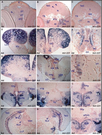
miR-221 expression in the zebrafish brain.
miR-221 shows spatially localized expression. It is strongly expressed in lateral hypothalamic areas (cells around the lateral recess, in the lateral torus and diffuse inferior lobe) and at high levels in caudal pallium and eminentia thalami. It is weakly expressed in a few rostral ventral telencephalic, thalamic, periventricular posterior tubercular, semicircular torus and rostral tectal cells (Table D). miR-221 expression is largely conserved in the adult telencephalic, hypothalamic, thalamic, posterior tubercular and semicircular torus nuclei (table I). However, there are some differences between the larval and adult miR-221 brain expression. miR-221 is expressed in adult preoptic area cells, isthmic nucleus, facial and vagal lobes, areas devoid of larval expression. miR-221 is also expressed in two cell populations in the adult tectal periventricular gray zone, area with few weakly stained cells in the larva.
miR-221 belongs to the same cluster as miR-222 and seems to largely share expression patterns but they also have subtle differences in transcript localisation. For instance, only miR-222 is expressed in the larval ventral intermediate hypothalamus, whereas only miR-221 is expressed in adult thalamic and pretectal nuclei, many adult migrated posterior tuberculum nuclei, larval and adult semicircular torus cells (tables D, I).

A. transverse section through the larval diencephalon showing weakly miR-221 expressing cells in the eminentia thalami (ET) and thalamic area (arrowhead, dorsal thalamus-DT, ventral thalamus-VT).
B. transverse section through the larval diencephalon and midbrain showing miR-221 strongly expressing cells at the level of the intermediate hypothalamus (Hi) in the diffuse nucleus of the hypothalamic inferior lobe (DIL) and the lateral torus (TLa) and weakly expressing cells in the optic tectum (arrowheads, TeO).
C. transverse section through the larval hypothalamus and midbrain showing miR-221 strongly expressing cells at the level of the intermediate hypothalamus (Hi) in the diffuse nucleus of the hypothalamic inferior lobe (DIL).
D. transverse section through the adult rostral telencephalon showing robust expression of miR-221 in the dorsal telencephalon/pallium (P) and weakly in the internal cellular layer of the olfactory bulb (ICL).
E. transverse section through the adult telencephalon showing miR-221 expressing cells in the ventral (Vv) and dorsal (Vd) nuclei of the ventral telencephalon (subpallium), medial (Dm), central (Dc), dorsal (Dd) and lateral (Dl) zones of the dorsal telencephalon.
F. transverse section through the adult preoptic area at the level of the optic chiasma (posterior to section E) showing miR-221 expressing cells in the suprachiasmatic nucleus (SC), preoptic posterior parvocellular (PPp) and magnocellular (PM) nuclei.
G. transverse section through the adult telencephalon at the level of anterior commissure (ac) showing miR-221 expressing cells in the supracommissural nucleus of the ventral telencephalon (Vs), medial (Dm), dorsal (Dd), lateral (Dl) and posterior (Dp) zones of the dorsal telencephalon.
H. transverse section through the adult diencephalon showing miR-221 expressing cells in the ventrolateral thalamic nucleus (VL) and the caudal telencephalon (Te).
I. Higher magnification of the section E at the level of the thalamus showing miR-221 expressing cells in the ventrolateral thalamic nucleus (VL).
J. transverse section through the adult hypothalamus showing miR-221 expressing cells in the ventral zone of periventricular hypothalamus (Hv), anterior tuberal nucleus (ATN), diffuse nucleus of the hypothalamic inferior lobe (DIL), nucleus of the paraventricular organ (nPVO), nucleus of periventricular posterior tuberculum (TPp) and medial preglomeral nucleus (PGm).
K. transverse section through the adult hypothalamus showing miR-221 expressing cells in the caudal zone of the periventricular hypothalamus (Hc), dorsal zone of the periventricular hypothalamus (Hd, at the level of lateral ventricular recess-lr) and diffuse nucleus of the hypothalamic inferior lobe (DIL).
L. transverse section through the adult hypothalamus showing miR-221 expressing cells in the ventral (Hv) and dorsal (Hd) zones of periventricular hypothalamus, anterior tuberal nucleus (ATN), lateral hypothalamic nucleus (LH), and nucleus of periventricular posterior tuberculum (TPp).
M. transverse section through the adult isthmus and caudal midbrain showing miR-221 strongly expressing cells in the tectal periventricular gray zone (pgz), central nucleus of semicircular torus (TSc, arrowheads), and weakly in the nucleus of lateral valvula (NLV, arrowhead).
N. transverse section through the adult isthmus and caudal midbrain showing miR-221 strongly expressing cells in the tectal periventricular gray zone (pgz) of the optic tectum (TeO), central nucleus of semicircular torus (TSc), isthmic nucleus (NI) and weakly in the nucleus of lateral valvula (NLV, arrowhead).
O. transverse section through the adult hypothalamus showing miR-221 expressing cells in the anterior tuberal nucleus (ATN), lateral hypothalamic nucleus (LH) and nucleus of the paraventricular organ (nPVO).

miR-222 expression in the zebrafish brain.
miR-222 expression is restricted to differentiating cells of the forebrain and midbrain and expression is largely conserved in these areas throughout life. In addition, miR-222 is de novo expressed in the adult facial and vagal lobes. In the larval brain, miR-222 is expressed in cells of all telencephalic areas apart from the lateral telencephalic area M4. In the diencephalon, miR-222 is expressed in the preoptic area, ventral thalamus and eminentia thalami, rostral and intermediate hypothalamic nuclei. More caudally, it is expressed in ventral and migrated posterior tubercular cells and a few scattered cells of the tectal periventricular gray zone (Table D). In the adult brain, miR-222 expression is conserved in the above areas with the exception of the thalamus where it is downregulated. In addition, miR-222 is expressed in the adult facial and vagal lobes although in the larva we did not observe expression in any hindbrain cells that could correspond to these adult areas (Table I).

A. transverse section through the larval telencephalon showing miR-222 expressing cells in the ventral (Sv) and dorsal (Sd) subpallium and pallium (P).
B. transverse section through the larval telencephalon (caudal to section A, at the level of the anterior commissure, arrowhead) showing miR-222 expressing cells in the preoptic area (Po), dorsal subpallium (Sd) and pallium (P).
C. transverse section through the larval caudal telencephalon and rostral diencephalon showing miR-222 expressing cells in the preoptic area (Po), eminentia thalami (ET), ventral thalamus (VT) and pallium (P).
D. transverse section through the larval hypothalamus and midbrain showing miR-222 expressing cells in the intermediate hypothalamus (Hi, ventral and at the level of lateral ventricular recess-lr), diffuse nucleus of the hypothalamic inferior lobe (DIL), lateral torus (TLa), ventral periventricular (PTv) and migrated posterior tubercular area (M2).
E. transverse section through the larval hindbrain at the level of the posterior lateral line ganglion (PLLG) where the medulla oblongata (MO) is devoid of miR-222 expression. The dorsal medulla oblongata at this level may give rise to cells of the facial and vagal lobes.
F. transverse section through the young adult caudal telencephalon at the level of the anterior commissure (ac) showing miR-222 expressing cells in the anterior parvocellular preoptic nucleus (PPa), endopeduncular nucleus (EN, part of eminentia thalami), supracommissural nucleus of the ventral telencephalon (Vs), medial (Dm) and lateral (Dl) zones of the dorsal telencephalon.
G. transverse section through the adult hypothalamus showing miR-222 expressing cells in the ventral zone of the periventricular hypothalamus (Hv), anterior tuberal nucleus (ATN), dorsal zone of the periventricular hypothalamus at the level of the lateral hypothalamic ventricular recess (Hd-lr), diffuse nucleus of the inferior hypothalamic lobe (DIL), lateral hypothalamic torus (TLa) and nucleus of the paraventricular organ (nPVO).
H. transverse section through the adult hypothalamus showing miR-222 expressing cells in the anterior tuberal nucleus (ATN), dorsal zone of the periventricular hypothalamus at the level of the lateral hypothalamic ventricular recess (Hd-lr), diffuse nucleus of the inferior hypothalamic lobe (DIL), lateral hypothalamic torus (TLa), nucleus of the paraventricular organ (nPVO) and posterior thalamic nucleus (Pt).
I. transverse section through the adult caudal hindbrain showing miR-222 expressing cells superficially lining the facial (LVII) and vagal (LX) lobes.
J. transverse section through the adult rostral telencephalon showing miR-222 expressing cells in the dorsal telencephalon/pallium (P) and internal cellular layer of olfactory bulb (ICL).
K. transverse section through the adult telencephalon showing miR-222 expressing cells in the ventral (Vv) and dorsal (Vd) nuclei of the ventral telencephalon (subpallium), medial (Dm), central (Dc), dorsal (Dd), lateral (Dl) and posterior (Dp) zones of the dorsal telencephalon.
L. transverse section through the adult telencephalon at the level of anterior commissure (ac) showing miR-222 expressing cells in the anterior parvocellular preoptic nucleus (PPa), dorsal part of the entopeduncular nucleus (ENd, part of the eminentia thalami), supracommissural/posterior nucleus of the ventral telencephalon (Vs), medial (Dm), dorsal (Dd) and lateral (Dl) zones of the dorsal telencephalon.

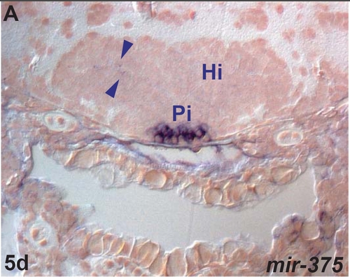
miR-375 expression in the zebrafish brain.
miR-375 shows cell type specific expression the pituitary, a few hypothalamic cells and cranial nerve ganglia (Table E).

A. transverse section through the embryonic intermediate hypothalamus (Hi) showing weak miR-375 expression in the hypothalamus (arrowheads) and strong expression in the pituitary (Pi).

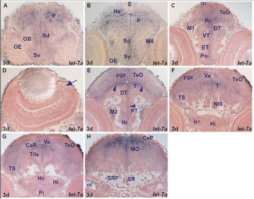
let-7a expression in the zebrafish brain.
let-7a, let-7b and let-7c are expressed in both proliferating and differentiating cells. let-7b and let-7c differ in their sequence in only one nucleotide located outside the seed region. They share similar regional expression in the larval brain with two differences: let-7b is expressed in the retinal ciliary marginal zone and pineal cells whereas let-7a and let-7c are absent (table A). let-7a, let-7b and let-7c mainly conserve their regional expression between larval and adult brain (tables A, F).
let-7a, let-7b and let-7c are expressed in many proliferating and differentiating cells of the larval fore-, mid- and hindbrain with the exception of some areas such as hypothalamic nuclei (caudal hypothalamus, diffuse nucleus of inferior lobe, lateral torus) interpeduncular nucleus, locus coereleus, raphe and reticular formation. We detected only minor differences at the regional level between larval and adult brain expression. For example, let-7b and let-7c are expressed in some adult but not larval hypothalamic lateral torus and superior raphe cells (tables A, F).

A. transverse section through the larval telencephalon showing let-7a expressing cells in the ventral (Sv) and dorsal (Sd) subpallium and pallium (P).
B. transverse section through the larval telencephalon and epithalamus showing let-7a expressing cells in the ventral (Sv) and dorsal (Sd) subpallium, pallium (P), migrated telencephalic area (M4) and habenula (Ha). Pineal cells (E) are devoid of expression.
C. transverse section through the larval diencephalon and rostral optic tectum showing let-7a expressing cells in the preoptic area (Po), eminentia thalami (ET), ventral thalamus (VT) and dorsal (DT) thalamus, periventricular (Pr) and migrated (M1) pretectum and optic tectum (TeO, including the tectal proliferative zone, m).
D. transverse section through the larval retina (dorsal to the right) devoid of let-7a expressing cells. The arrow points at the ciliary marginal zone.
E. transverse section through the larval diencephalon and midbrain showing mainly periventricular (arrowheads) let-7a expressing cells in the rostral hypothalamus (Hr), periventricular (PT) and migrated (M2) posterior tuberculum, dorsal thalamus (DT), tegmentum (T) and tectal periventricular gray zone (pgz).
F. transverse section through the larval hypothalamus, midbrain and rostral hindbrain showing let-7a expressing cells in the intermediate hypothalamus (Hi, area of the periventricular hypothalamic recess-lr), semicircular torus (TS), tegmentum (T), tectal periventricular gray zone (pgz) and cerebellar valvula (Va).
G. transverse section through the larval hypothalamus, midbrain and rostral hindbrain showing let-7a expressing cells in the intermediate (Hi) and caudal (Hc) hypothalamus, semicircular torus (TS), tegmentum/isthmic area (T/Is), periventricular gray zone (pgz) of the optic tectum (TeO), cerebellar valvula (Va) and cerebellar plate (CeP).
H. transverse section through the larval hindbrain at the level of the otic capsule (ot) showing let-7a expressing cells in the medulla oblongata (MO).

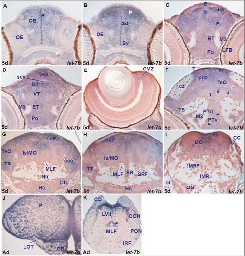
let-7b expression in the zebrafish brain.
let-7a, let-7b and let-7c are expressed in both proliferating and differentiating cells. let-7b and let-7c differ in their sequence in only one nucleotide located outside the seed region. They share similar regional expression in the larval brain with two differences: let-7b is expressed in the retinal ciliary marginal zone and pineal cells whereas let-7a and let-7c are absent (table A). let-7a, let-7b and let-7c mainly conserve their regional expression between larval and adult brain (tables A, F).
let-7a, let-7b and let-7c are expressed in many proliferating and differentiating cells of the larval fore-, mid- and hindbrain with the exception of some areas such as hypothalamic nuclei (caudal hypothalamus, diffuse nucleus of inferior lobe, lateral torus) interpeduncular nucleus, locus coereleus, raphe and reticular formation. We detected only minor differences at the regional level between larval and adult brain expression. For example, let-7b and let-7c are expressed in some adult but not larval hypothalamic lateral torus and superior raphe cells (tables A, F).

A. transverse section through the larval rostral telencephalon showing let-7b expressing cells in the olfactory bulb (OB).
B. transverse section through the larval telencephalon showing let-7b expressing cells in the ventral (Sv) and dorsal (Sd) subpallium and pallium (P).
C. transverse section through the larval rostral diencephalon showing let-7b expressing cells in the preoptic area (Po), eminentia thalami (ET), migrated eminentia thalami (M3), pallium (P), habenula (Ha) and epiphysis (E).
D. transverse section through the larval diencephalon and rostral optic tectum showing let-7b expressing cells in the preoptic area (Po), eminentia thalami (ET), migrated eminentia thalami (M3), ventral thalamus (VT), dorsal thalamus (DT), and optic tectum (TeO).
E. transverse section through the larval retina (dorsal to the right) showing let-7b expressing cells in the ciliary marginal zone (CMZ).
F. transverse section through the larval diencephalon and midbrain showing mainly periventricular (arrowheads) let-7b expressing cells in the dorsal (PTd) and ventral (PTv) periventricular and lateral migrated (M2) posterior tuberculum, tegmentum (T), semicircular torus (TS), longitudinal torus (TL), tectal proliferative (m) and periventricular gray (pgz) zones.
G. transverse section through the larval caudal midbrain and rostral hindbrain showing let-7b expressing cells in the semicircular torus (TS), optic tectum (TeO), isthmic/medulla oblongata area (Is/MO) and cerebellar plate (CeP).
H. transverse section through the larval rostral hindbrain showing let-7b expressing cells in the semicircular torus (TS), cerebellar plate (CeP) and isthmic/medulla oblongata area (Is/MO).
I. transverse section through the larval hindbrain at the level of the otic capsule showing let-7b expressing cells in the medulla oblongata (MO) and rhombic lip (RL).
J. transverse section through the adult rostral telencephalon showing let-7b expressing cells in the dorsal telencephalon/pallium (P) and olfactory bulb (OB).
K. transverse section through the adult caudal hindbrain showing let-7b expressing cells in the inferior reticular formation (IRF), posterior octaval (PON) and caudal octavolateral (CON) areas, facial lobe (LVII) and around the rhomboncephalic ventricle (arrowhead).

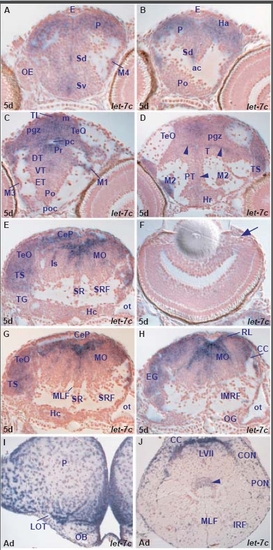
let-7c expression in the zebrafish brain.
let-7a, let-7b and let-7c are expressed in both proliferating and differentiating cells. let-7b and let-7c differ in their sequence in only one nucleotide located outside the seed region. They share similar regional expression in the larval brain with two differences: let-7b is expressed in the retinal ciliary marginal zone and pineal cells whereas let-7a and let-7c are absent (table A). let-7a, let-7b and let-7c mainly conserve their regional expression between larval and adult brain (tables A, F).
let-7a, let-7b and let-7c are expressed in many proliferating and differentiating cells of the larval fore-, mid- and hindbrain with the exception of some areas such as hypothalamic nuclei (caudal hypothalamus, diffuse nucleus of inferior lobe, lateral torus) interpeduncular nucleus, locus coereleus, raphe and reticular formation. We detected only minor differences at the regional level between larval and adult brain expression. For example, let-7b and let-7c are expressed in some adult but not larval hypothalamic lateral torus and superior raphe cells (tables A, F).

A. transverse section through the larval telencephalon showing let-7c expressing cells in the ventral (Sv) and dorsal (Sd) subpallium and pallium (P).
B. transverse section through the larval caudal telencephalon and epithalamus showing let-7c expressing cells in the dorsal subpallium (Sd), pallium (P) and habenula (Ha).
C. transverse section through the larval diencephalon and optic tectum showing let-7c expressing cells in the ventral thalamus (VT), dorsal thalamus (DT), periventricular (Pr) and migrated (M1) pretectum and optic tectum (TeO, proliferative zone-m, periventricular gray zone-pgz, longitudinal torus-TL).
D. transverse section through the larval diencephalon and midbrain showing mainly periventricular (arrowheads) let-7c expressing cells in the periventricular (PT) and lateral migrated (M2) posterior tuberculum, tegmentum (T), semicircular torus (TS) and tectal periventricular gray zone (pgz).
E. oblique transverse section through the larval caudal hypothalamus, midbrain and hindbrain showing let-7c expressing cells in the semicircular torus (TS), optic tectum (TeO), isthmic area (Is), cerebellar plate (CeP), and medulla oblongata (MO).
F. transverse section through the larval retina (dorsal to the right) devoid of let-7c expressing cells. The arrow points at the ciliary marginal zone.
G. oblique transverse section through the larval hindbrain at the level of the otic capsule (caudal to section E) showing let-7c expressing cells in the semicircular torus (TS), optic tectum (TeO), granular cerebellar eminence (EG), cerebellar plate (CeP) and medulla oblongata (MO).
H. oblique transverse section through the larval hindbrain at the level of the octaval ganglion (caudal to section G) showing let-7c expressing cells in the granular cerebellar eminence (EG), medulla oblongata (MO) and rhombic lip (RL).
I. transverse section through the adult rostral telencephalon showing let-7c expressing cells in the dorsal telencephalon/pallium (P) and olfactory bulb (OB).
J. transverse section through the adult caudal hindbrain showing let-7c expressing cells in the inferior reticular formation (IRF), posterior octaval (PON) and caudal octavolateral (CON) nuclei and facial lobe (LVII).

Other miRNAs expressed in the zebrafish brain.
In addition to the 21 described above, we have examined the expression of seventeen additional miRNAs in the zebrafish brain. In this figure and following tables (Additional file AF27) we show examples of their expression.

A. transverse section through the larval rostral telencephalon showing miR-139 expressing cells in the olfactory epithelium (OE). miR-139 is widely expressed in differentiated cells in the zebrafish brain (see also panel D).
B. transverse section through the adult olfactory bulb showing miR-187 expressing cells in glomerular (GL) and internal cellular (ICL) layers. miR-187 is widely expressed in differentiated cells in the adult zebrafish brain.
C. transverse section through the embryonic rostral diencephalon showing miR-454a expressing cells in the preoptic area (Po), eminentia thalami (ET), migrated eminentia thalami (M3), habenula (Ha), and retina. The arrow points at the ciliary marginal zone. miR-454a is almost ubiquitously expressed in the larval zebrafish brain.
D. transverse section through the adult hypothalamus showing miR-139 expressing cells in ventral (Hv) and dorsal (Hd) zones of periventricular hypothalamus, anterior tuberal nucleus (ATN), lateral hypothalamic nucleus (LH), posterior tuberal nucleus (PTN), lateral torus (TLa) and lateral preglomerular nucleus (PGl). miR-139 is widely expressed in differentiated cells in the zebrafish brain.
E. transverse section through the larval retina showing miR-132 expressing cells in the ciliary marginal zone (CMZ, arrow), inner nuclear layer (INL) and ganglion (GCL) cell layers. miR-132 is widely expressed in the zebrafish brain.
F. transverse section through the larval retina showing miR-125b expressing cells in the ciliary marginal zone (CMZ, arrow), inner (INL) and ganglion (GCL) cell layers. miR-125b is almost ubiquitously expressed in the zebrafish brain.
G. transverse section through the adult hypothalamus showing miR-132 expressing cells in dorsal zone of periventricular hypothalamus (Hd), anterior tuberal nucleus (ATN), lateral hypothalamic nucleus (LH), posterior tuberal nucleus (PTN), medial preglomerular nucleus (PGm) and posterior thalamic nucleus (Pt). miR-132 is widely expressed in differentiated cells in the zebrafish brain (see also panel E).
H. transverse section through the adult preoptic area showing miR-98 expressing cells in the suprachiasmatic nucleus (SC) and parvocellular posterior preoptic nucleus (PPp). miR-98 is widely expressed in the adult zebrafish brain.
I. transverse section through the adult preoptic area showing miR-34b expressing cells lining and lateral to the ventricle in the anterior parvocellular preoptic nucleus (PPa). miR-34b is mainly expressed in periventricular cells of the adult zebrafish brain (see also panel L).
J. transverse section through the larval diencephalon and optic tectum (TeO) showing miR-125a expressing cells in the ventral (VT) and dorsal (DT) thalamus and the periventricular gray zone (pgz) of the TeO. miR-125a is widely expressed in differentiated cells of the zebrafish brain.
K. transverse section through the adult dorsal diencephalon showing miR-100 expressing cells in the habenular nuclei (dorsal-Had, ventral-Hav), ventromedial thalamic nucleus (VM) and optic tectum (TeO). miR-100 is widely expressed in the zebrafish brain.
L. transverse section through the adult thalamic area showing miR-34b expressing cells lining the ventricle in the ventromedial thalamic nucleus (VM). miR-34b is mainly expressed in periventricular cells of the adult zebrafish brain (see also panel I).
M. transverse section through the larval midbrain showing miR-99 expressing cells in the periventricular gray zone (pgz) of the optic tectum (TeO), tegmentum (T), semicircular torus (TS) and intermediate hypothalamus (Hi). miR-99 is widely expressed in periventricular and differentiating cells of the larval zebrafish brain.
N. transverse section through the adult isthmus showing miR-103 expressing cells in the central gray (GC), superior raphe (SR), isthmic nucleus (NI), nucleus of lateral valvula (NLV) and diffuse nucleus of inferior hypothalamic lobe (DIL). miR-103 is widely expressed in the zebrafish brain.
O. transverse section through the adult cerebellum showing miR-16 expressing cells in the cerebellar granular layer (CeGL). miR-16 is widely expressed in the zebrafish brain.

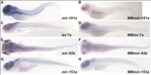
Mismatch test for let-7a, miR-92b, miR-153a and miR-181a.
To test the ability of LNA probes to discriminate between different members within a miRNA family, we performed in situ hybridization with single and double mismatch probes for let-7a, miR-92b, miR-153a and miR-181a. In parallel we performed in situ hybridization with the fully matching LNA probes. All probes were labeled and hybridized in parallel.

A,C,E,G: show in situ hybridization signal for full matching LNA probes miR-181a, let-7a, miR-92b and miR-153a, respectively.
B,D,F,H: show in situ hybridization signal for a double mismatch miR-181a probe (MMmiR-181a) and single mismatch probes let-7a, miR-92b and miR-153a (MMlet-7a, MMmiR-92b and MMmiR-153a), respectively.
Although there is a strong reduction in hybridization signal with the single mismatch probes (MMlet-7a, MMmiR-92b and MMmiR-153a), hybridization was only fully eliminated by the double mismatch probe MMmiR-181a. Thus, LNA probes may not be able to fully discriminate between miRNA family members that differ at a single position.

Acknowledgments
This image is the copyrighted work of the attributed author or publisher, and ZFIN has permission only to display this image to its users. Additional permissions should be obtained from the applicable author or publisher of the image.
Open Access.
Full text @ Genome Biol.