|
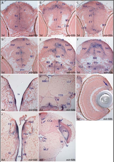
miR-92b expression in the zebrafish brain.
miR-92b is predominantly expressed in periventricular and adjacent cells corresponding to proliferative zones throughout the 5dpf larval brain (table A). It is also expressed in the retinal ciliary marginal zone. miR-92b expression is conserved in the same cell types in the adult brain (table F). A. transverse section through the larval telencephalon showing miR-92b expressing cells in the ventral (Sv) and dorsal (Sd) subpallium, pallium (P) and epiphysis (E).
B. transverse section through the caudal telencephalon and rostral diencephalon showing miR-92b strongly expressing cells lining and close to the ventricle between the pallium (P) and epithalamus (habenula-Ha and epiphysis-E).
C. transverse section through the larval diencephalon and rostral optic tectum showing miR-92b expressing cells lining and close to the ventricle in the preoptic area, (Po), eminentia thalami (ET), ventral thalamus (VT), dorsal thalamus (DT) and the optic tectum (TeO).
D. transverse section through the larval diencephalon and optic tectum showing miR-92b expressing cells lining and close to the ventricle in the preoptic area (Po), ventral thalamus (VT), dorsal thalamus (DT), periventricular pretectum (Pr) and the proliferative zone of the optic tectum (m).
E. oblique transverse section through the larval midbrain, hypothalamus and rostral isthmus showing miR-92b expressing cells in the intermediate hypothalamus (Hi) around the lateral ventricular recess (lr, arrowheads), ventral posterior periventricular tuberculum (PTv), lining and close to the ventricle of tegmentum (T) and semicircular torus (TS), in the cerebellar valvula (Va) and proliferative zone of the optic tectum (m).
F. transverse section through the larval midbrain, hypothalamus and rostral isthmus showing miR-92b expressing cells in the intermediate hypothalamus (Hi) around the lateral ventricular ventricular recess (lr), lining and close to the ventricle in the tegmentum and isthmus (T/Is), semicircular torus (TS), in the cerebellar valvula (Va), cerebellar plate (CeP) and proliferative zone of the optic tectum (m).
G. transverse section through the adult telencephalon showing miR-92b expressing cells lining the telencephalic ventricle (tv) in the subpallium (dorsal nucleus of the ventral telencephalon,Vd) and the pallium (medial nucleus of the dorsal telencephalic area, Dm).
H. transverse section through the adult midbrain and hypothalamus showing miR-92b expressing cells lining/close to the ventricle in the dorsal (Hd-lr) and caudal (Hc) zones of the periventricular hypothalamus, mammillary body (CM) and lateral torus (TLa).
I. transverse section through the larval retina showing miR-92b expressing cells in the ciliary marginal zone (arrow).
J. transverse section through the adult dorsal diencephalon showing miR-92b expressing cells lining/close to the ventricle in the dorsal (Had) and ventral (Hav) habenular nuclei, anterior (A) and ventromedial (VM) thalamic nuclei.
K. transverse section through the adult cerebellum, isthmus and caudal midbrain. Arrows point at miR-92b expressing cells lining/close to the ventricle in the central gray (GC), optic tectum (TeO), central semicircular torus (TSc) and cerebellum (CCe).
|