
|
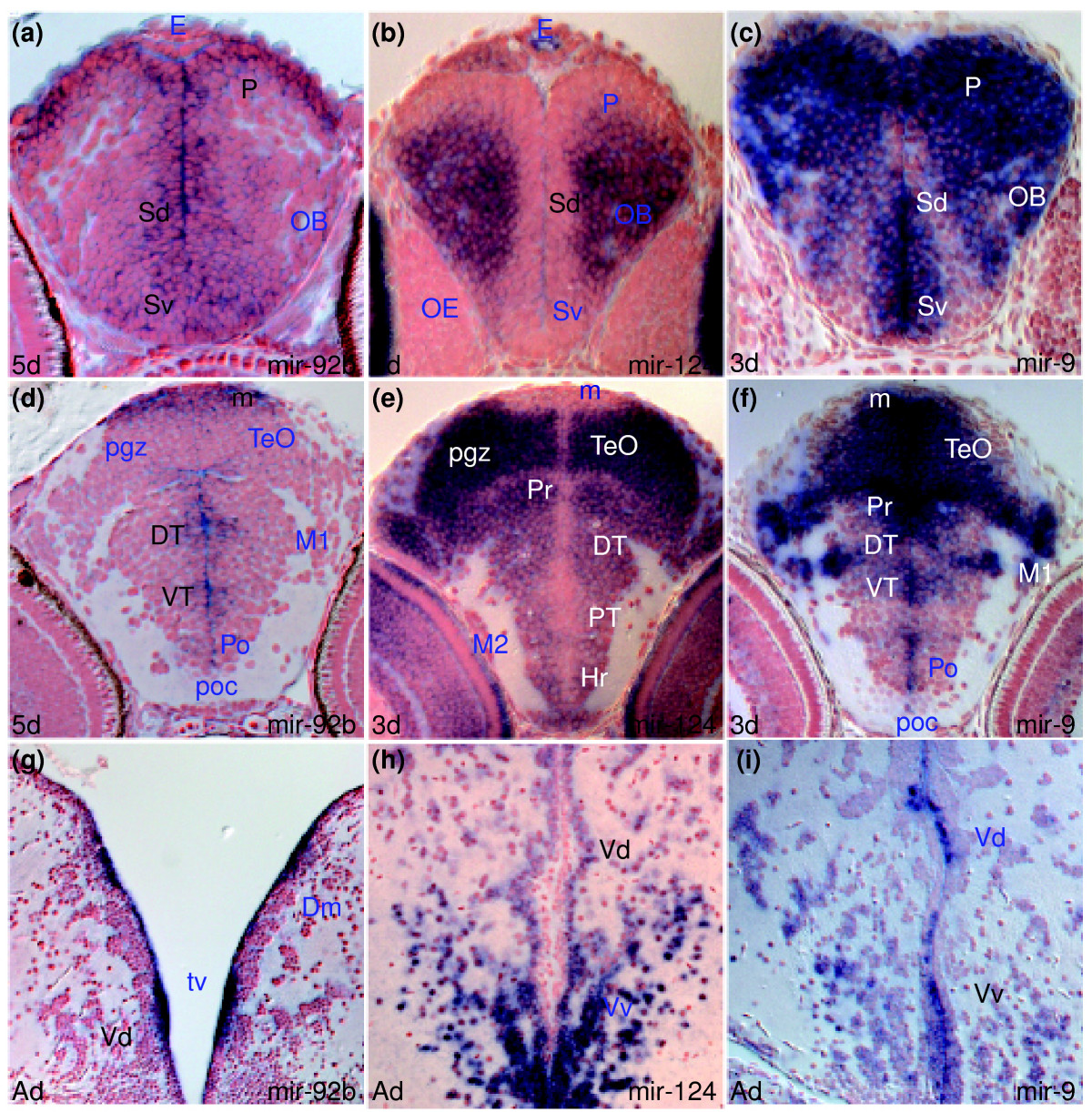
Fig. 1 miRNAs expressed in proliferating and/or differentiating cells in the developing and adult zebrafish brain. In this and other figures, unless otherwise mentioned, sections are transverse with dorsal on the top, stage is shown bottom left and miRNA analyzed by in situ hybridization bottom right, in situ staining is in blue and cell nuclei are visualized with nuclear red counterstaining. Abbreviations used in the Results section of the text are denoted in black. For other abbreviations, see Additional data file 26. (a,d,g) miR-92b expression in periventricular and adjacent cells of the telencephalon (a,g), diencephalon and optic tectum (d). (b,e,h) miR-124 expression in differentiating cells in the telencephalon (b,h), diencephalon and optic tectum (e). (c,f,i) miR-9 expression in periventricular/proliferating and differentiating cells of the telencephalon (c,i), diencephalon and optic tectum (f).