
- Title
-
lunatic fringe regulates delta-notch induction of hypochord in zebrafish
- Authors
- Appel, B., Marasco, P., McClung, L.E., and Latimer, A.J.
- Source
- Full text @ Dev. Dyn.
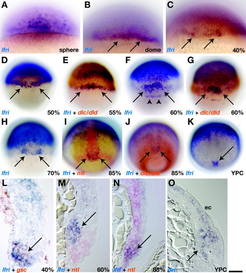
Dorsal margin cells transiently express lfng during gastrulation. A,B: Lateral views; C-K: dorsal views; and L-O: sagittal sections of blastula and gastrula stage embryos processed for in situ RNA hybridization. A: Sphere stage (4 hours postfertilization [hpf]). lfng expression was punctate throughout the blastoderm. B: Dome stage (4.3 hpf). Arrows indicate lfng+ cells near the embryonic margin. C,D: Arrows indicate lfng+ cells concentrated at the dorsal margin at 40% epiboly (5 hpf) and 50% epiboly (5.3 hpf). E: At 55% epiboly, lfng+ cells occupied a more narrow area of the dorsal margin (arrows) and appeared to be adjacent to cells that expressed dlc and dld (red). F: At 60% epiboly (6 hpf). Arrows indicate lfng+ cells located just above the dorsal margin, and arrowheads mark lfng+ dorsal forerunner cells. G: Dorsal lfng+ cells appeared to be adjacent to dlc- and dld-expressing cells. H: At 70% epiboly (8 hpf). Arrows mark lfng+ cells at the dorsal margin. I,J: At 85% epiboly (8.6 hpf). Embryos continued to express lfng at the dorsal margin (arrows), in cells that also expressed ntl (I) and that were adjacent to dlc/dld+ cells (J). K: At yolk plug closure (YPC) stage (10 hpf). Arrow indicates lfng expression at dorsal margin. L: At 40% epiboly. Dorsal margin cells (arrow) coexpressed gsc (red) and lfng (blue). M: At 60% epiboly lfng+ cells (blue, arrow) occupied dorsal mesendoderm, just anterior to ntl+ cells (red). N: At 85% epiboly. Dorsal margin cells (arrow) coexpressed lfng (blue) and ntl (red). O: YPC stage. lfng+ cells were deep in the nascent tail bud (arrow) and in ectoderm (ec). Scale bar = 80 μm in A-C, 160 μm in D-K, 40 μm in L-O. |
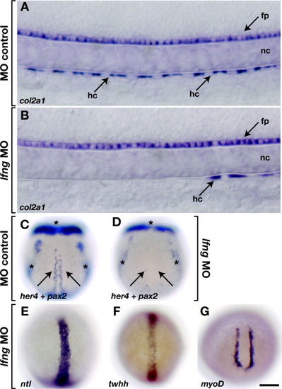
lfng is required for hypochord development. A: Lateral view, dorsal up, of 24 hours postfertilization (hpf) embryo injected with lfng 4 ng of mismatch control morpholino oligonucleotides (MO). Floor plate (fp), notochord (nc), and hypochord (hc) developed normally. B: A 24 hpf embryo injected with approximately 4 ng of lfng MO had a deficit of hypochord cells. Floor plate and notochord appeared normal. C: Dorsal view, anterior up, of yolk plug closure (YPC) stage embryo injected with standard control MO and probed for her4 expression, which marks presumptive hypochord precursors (arrows), and pax2, to provide an internal hybridization control (asterisks). D: her4 expression was absent (arrows) in embryo injected with approximately 4 ng of lfng MO, whereas pax2 expression was normal (asterisks). E-G: ntl, twhh, and myoD expression in lfng MO injected embryos. Scale bar in G = 20 μm in A,B, 160 μm in C-G. |
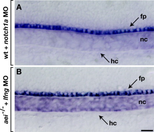
lfng potentiates Delta-Notch signaling in hypochord development. Lateral views, dorsal up, of 26-28 hours postfertilization embryos. A: Wild-type embryo injected with notch1a morpholino oligonucleotides (MO). Hypochord (hc) was absent, whereas floor plate (fp) and notochord (nc) appeared normal. B: aei-/- embryo injected with approximately 1 ng of lfng MO. Scale bar = 20 μm. EXPRESSION / LABELING:
|

Unillustrated author statements |