
|
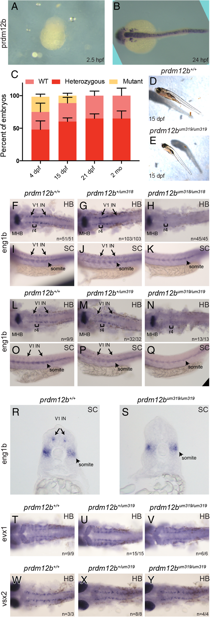
Fig. 2
prdm12b germ line mutants lack eng1b expression in the p1 domain. a, b. prdm12b is not maternally deposited. In situ hybridization detects prdm12b expression at 24hpf (b), but not at 2.5hpf (a), in wildtype embryos. c. Bar chart depicting the frequency of each genotype at various time points in broods from crosses of prdm12b heterozygous animals. Error bars indicate ±S. E. (n = 3). dpf = days post fertilization, mo = months. d, e. Morphology of 15dpf prdm12b+/+ (d) and prdm12bum319/um319 (e) fish. f-s. eng1b expression in 24hpf embryos from crosses of prdm12b+/um318 heterozygotes (f-k), or prdm12b+/um319 heterozygotes (l-s). Numbers in each panel indicate the fraction of animals with the specified phenotype. t, u. evx1expression in 24hpf embryos from a cross of prdm12b+/um319 heterozygotes. v, w. vsx2expression in 24hpf embryos from a cross of prdm12b+/um319 heterozygotes. Embryos are shown in dorsal (f-h, l-n, t-y) or lateral (i-k, o-q) view with anterior to the left, or in cross section (r, s) with dorsal to the top. Brackets indicate r4, arrows mark V1 interneurons and arrowheads mark somites. MHB = midbrain–hindbrain boundary, HB = hindbrain and SC = spinal cord