
Image
|
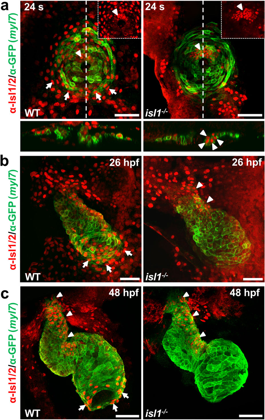
Figure Caption
Fig. 1
Residual Isl1/2 positive cells in isl1−/− zebrafish hearts.
(a–c) Confocal images of wild-type sibling and Tg(myl7:EGFP-HsHRAS)s883 isl1−/− embryos stained with anti-GFP and anti-Isl1/2 antibodies at 24 somites (a), 26 hpf (b) and 48 hpf (c). Arrows point to Isl1+ cells at the periphery of the cone (a) or Isl1+ cardiomyocytes at the venous pole of the atrium (b,c), arrowheads point to residual Isl1/2+ cardiomyocytes in the future ventricle (a) or the inner curvature of the ventricle and the outflow pole (b,c). Scale bars, 50 μm.
Figure Data
Acknowledgments
This image is the copyrighted work of the attributed author or publisher, and
ZFIN has permission only to display this image to its users.
Additional permissions should be obtained from the applicable author or publisher of the image.
Full text @ Sci. Rep.