
Fig. 5
|
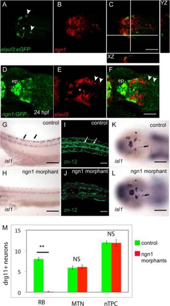
Fig. 5
Ngn1 function is required for RB neuron development, but not for MTN development. A–C: Dorsal views of the brains of 24-hpf Tg[elavl3:egfp] embryos processed by fluorescent in situ for ngn1 and labelled with anti-GFP. Orthogonal views at the level of MTN neurons show there is no co-localisation of ngn1 and EGFP in nTPC (asterisk) or MTN neurons (arrowheads, C). D–F: A comparison of huc/elavl3 expression to GFP in the brain of a 24-hpf Tg[-8.4neurog1:GFP] transgenic larvae does not reveal co-localisation in nTPC or MTN neurons, although some epiphyseal neurons (ep) do show co-expression. Uninjected (G,I,K) or embryos injected with ngn1 morpholino (H,J,L), then fixed at 24 hpf. Lateral views of the spinal cord reveal that isl1 expression in absent from RB neurons in ngn1 morphants (G,H). A dorsal view of the spinal cord labelled with zn-12 reveals a loss of RB neurons (arrows) and reticulospinal tract axons (I,J). Dorsal views of the head reveal that isl1 expressing MTN and nTPC neurons appear to be unaffected in ngn1 morphants (K,L). M: Quantification of RB, MTN,and nTPC neurons expressing drg11 reveals that ngn1 loss of function results in a dramatic reduction of RB neurons (**P<0.01), but does not result in a significant change (NS) of MTN or nTPC neuron number.