
|
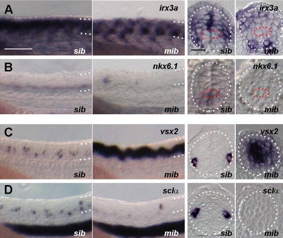
Fig. 1 Mib-mediated Notch signaling regulates both p2 progenitor maintenance and the V2a/V2b cell fates. (A, B) p2 progenitors in the ventricular zone were reduced in mib mutants. irx3a (A) or nkx6.1 (B) Expression in sibling control (sib) or mib mutants (mib). Increased ventral irx3a-expressing cells in mib mutants are primary motoneurons. (C, D) V2a was increased and expanded into the ventricular zone due to precocious differentiation and V2b was reduced in mib mutants. V2a and V2b cells were detected by vsx2 (C) and sclα (D), respectively in sibling control (sib) or mib mutants (mib). Left two panels: side views of embryos at 24 hpf with anterior to the left, dorsal up. Dorsal and ventral borders of the neural tubes are shown by the dotted lines. Bar scale: 100 µm. Right two panels: transverse sections through the trunk region in embryos at 24 hpf. p2 Progenitor domains within the neural tubes (circled by white dotted lines) are indicated by red dashed outlines. Bar scale: 20 µm.
Reprinted from Developmental Biology, 391(2), Okigawa, S., Mizoguchi, T., Okano, M., Tanaka, H., Isoda, M., Jiang, Y.J., Suster, M., Higashijima, S.I., Kawakami, K., Itoh, M., Different combinations of Notch ligands and receptors regulate V2 interneuron progenitor proliferation and V2a/V2b cell fate determination, 196-206, Copyright (2014) with permission from Elsevier. Full text @ Dev. Biol.