
Fig. 1
|
Fig. 1
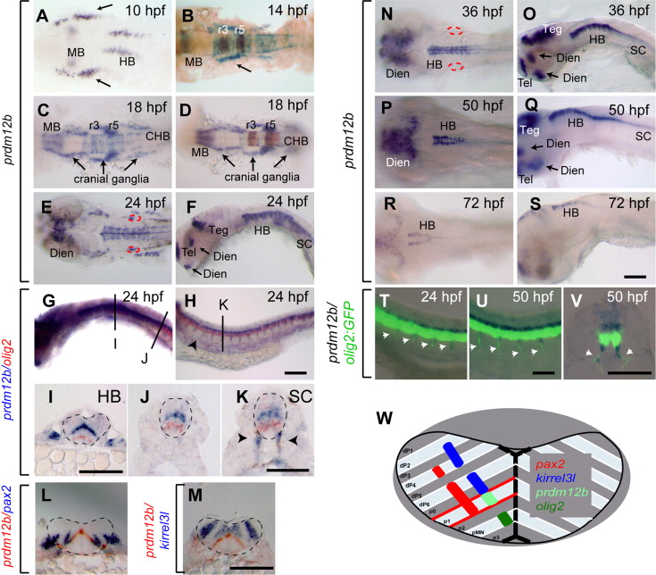
prdm12b is expressed in the p1 domain. (A–V). Expression of prdm12b by itself (A, C, E, F, N–S) and together with krox20 (B, D), olig2 (G–K), pax2 (L), kirrel3l (M) or olig2:egfp (T–V) in wild type embryos. Embryos are shown in dorsal (A–E, N, P, R) or lateral (F–H, O, Q, S–U) view with anterior to the left, or in cross section (I–M) with dorsal to the top. (W) Diagram of hindbrain gene expression. Scale bars are 100 μm, except (J–K, V) that are 50 µm. The otic vesicles and the neural tube are outline by red and black dashed lines, respectively. Arrows indicate cranial ganglia (A–D), or forebrain/midbrain structures (F, O, Q). Arrowheads mark motor axons (H, K, T–V). MB – midbrain, HB – hindbrain, CHB – caudal hindbrain, Teg – tegmentum, Dien – diencephalon, Tel – telencephalon, and r – rhombomere.
Reprinted from Developmental Biology, 390, Zannino, D.A., Downes, G.B., Sagerström, C.G., prdm12b specifies the p1 progenitor domain and reveals a role for V1 interneurons in swim movements, 247-60, Copyright (2014) with permission from Elsevier. Full text @ Dev. Biol.